Review
. 2018 May 25;8(35):19369-19380.
doi: 10.1039/c8ra02221f.

Graphitic-phase carbon nitride-based electrochemiluminescence sensing analyses: recent advances and perspectives

Affiliations
Review

Graphitic-phase carbon nitride-based electrochemiluminescence sensing analyses: recent advances and perspectives

Jingjing Jiang et al. RSC Adv. .

Abstract

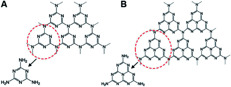
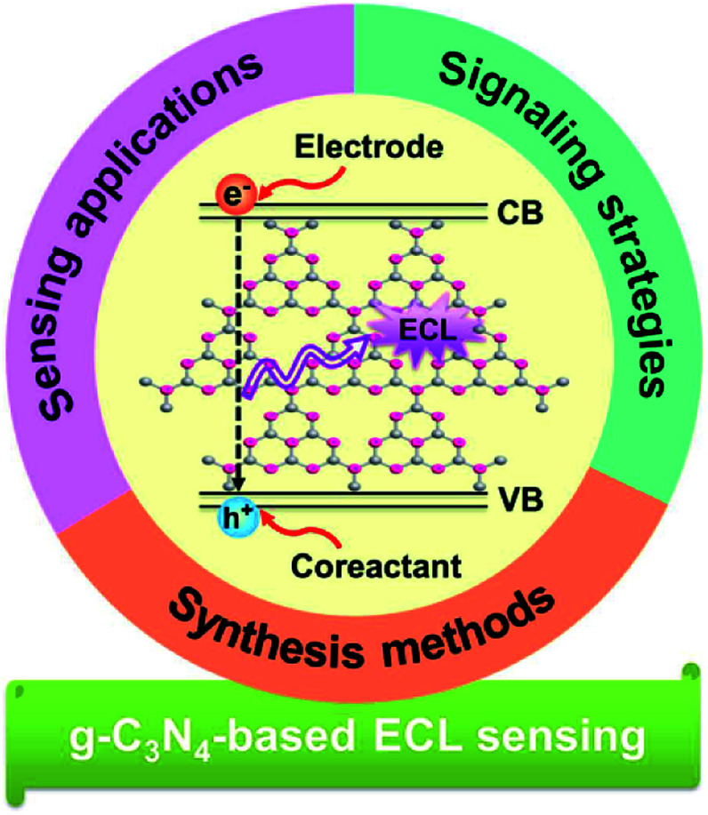
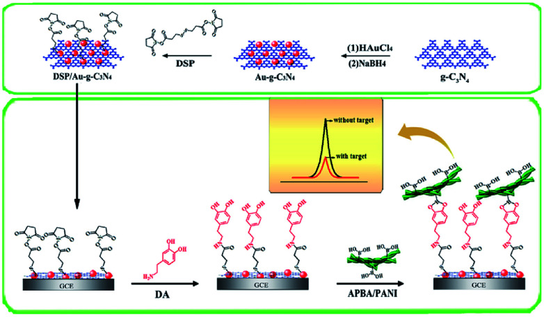
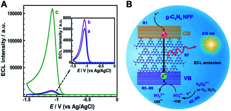
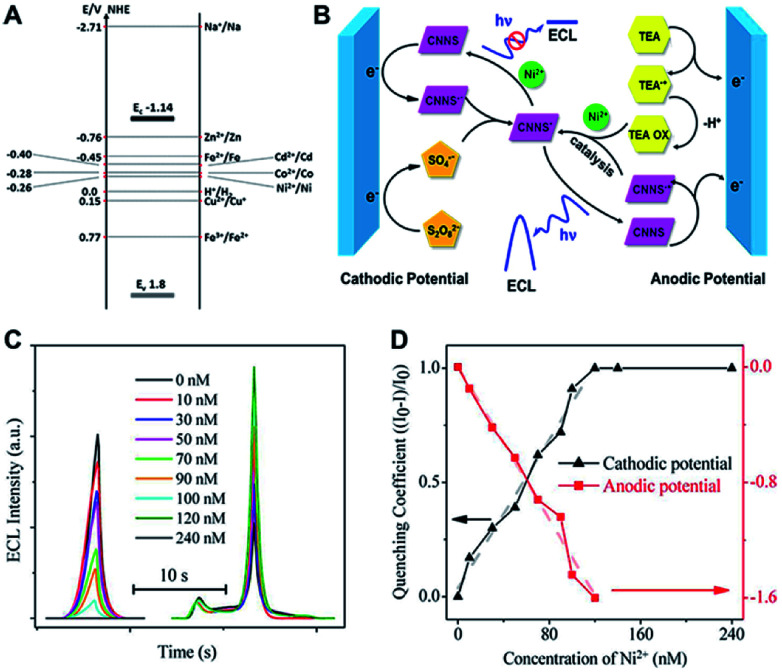
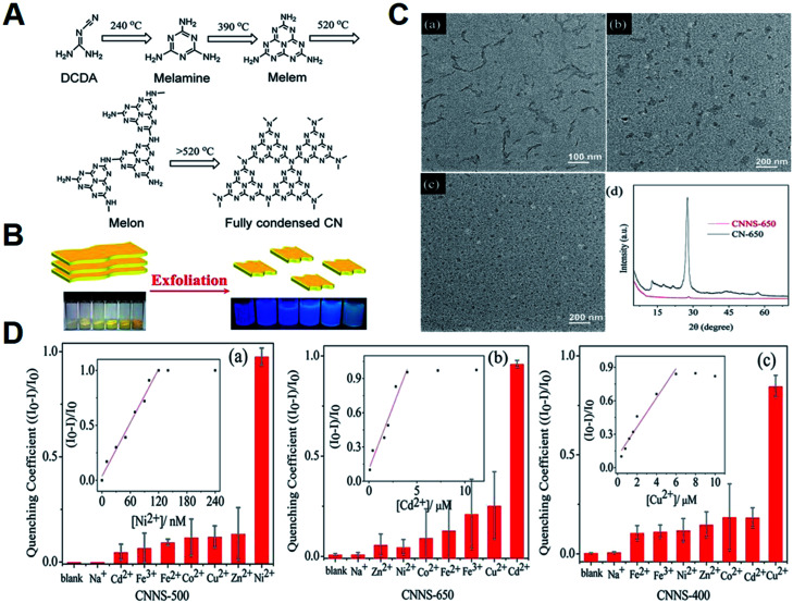
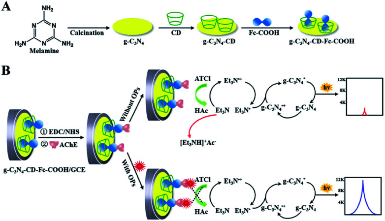
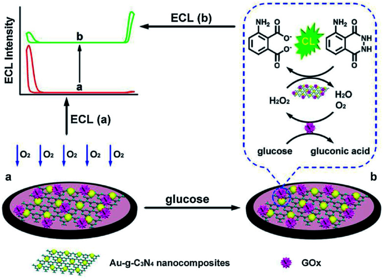
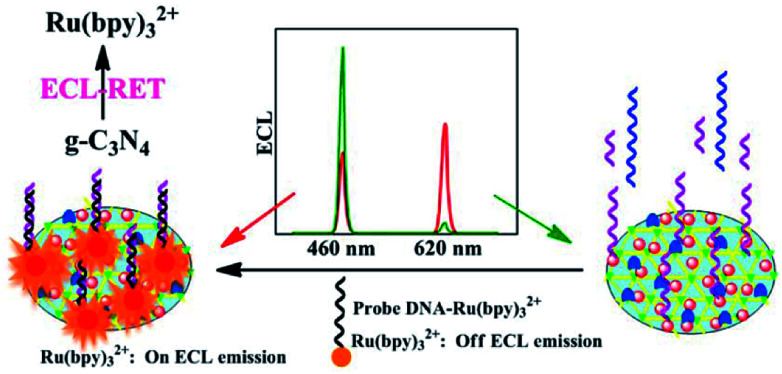
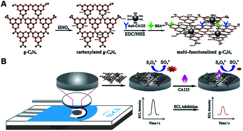
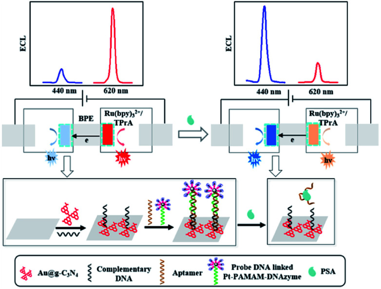
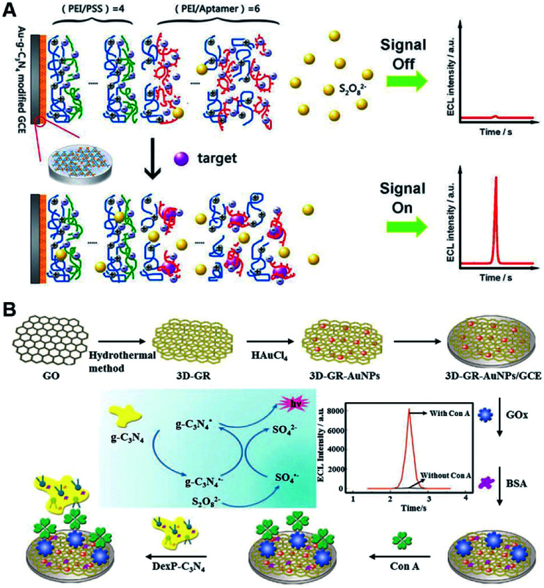
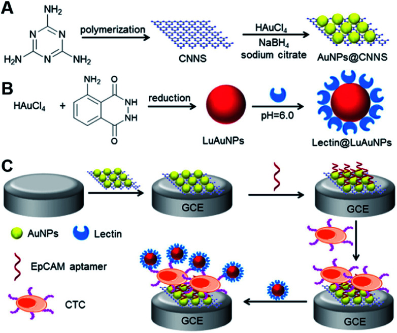
Graphitic-phase carbon nitride (g-C3N4) materials are important polymeric and metal-free semiconductors, and have attracted extensive attention as emerging electrochemiluminescence (ECL) emitters due to their wonderful optical and electronic properties. The g-C3N4-based ECL sensing analysis, as a research hotspot in analytical chemistry, offers an exquisite pathway to monitor target analytes with the advantages of low background signal, high sensitivity, desirable controllability, and simple instrumentation. Herein, we briefly describe the current research status of g-C3N4-based ECL assays along with versatile signaling strategies, introduce the preparation methods and ECL emission mechanisms of g-C3N4-dependent emitters, summarize their ECL sensing applications from 2012 to now, highlighting with special examples of metal ion and small molecule detection, nucleic acid bioanalysis, immunoassay, protein sensing, and cell-related determination. Finally, the prospects and challenges for future work are also explored to design more advanced ECL biosensors.


Conflict of interest statement

There are no conflicts to declare.

Figures

Fig. 1
Fig. 1. (A) Triazine and (B) tri-s-triazine as the basic structural units of g-C3N4. Reproduced from ref. 34 with permission from The American Chemical Society.
Fig. 2
Fig. 2. Overview of g-C3N4-based materials for ECL sensing analysis.
Fig. 3
Fig. 3. The preparation of DSP-decorated Au–g-C3N4 for highly selective ECL detection of DA based on dual-molecular recognition and PANI quenching. Reproduced from ref. 54 with permission from Elsevier.
Fig. 4
Fig. 4. (A) The ECL responses of g-C3N4 NFF modified GCE in 0.1 M (a) air-saturated PBS of pH 7.0, nitrogen-saturated PBS of pH 7.0 containing (b) 0.258 mM H2O2 and (c) 0.258 mM K2S2O8, and (B) the cathodic ECL emission mechanism for g-C3N4 NFF/R-O coreactant systems. Reproduced from ref. 63 with permission from The Royal Society of Chemistry.
Fig. 5
Fig. 5. (A) The electronic band structure of CNNS and oxidation/reduction potentials of different metal ions; (B) the proposed ECL reaction pathways of CNNS in the presence of Ni2+; (C) ECL intensity of CNNS-modified GCE with different Ni2+ concentrations in the presence of K2S2O8 at cathodic potential range and TEA at anodic potential range; and (D) the linear fitting of cathodic and anodic ECL quenching coefficients. Reproduced from ref. 68 with permission from The American Chemical Society.
Fig. 6
Fig. 6. (A) The typical reaction path for the formation of full condensed CN; (B) the process of exfoliating CN-T (T = 400, 450, 500, 550, 600, 650); (C) TEM of (a) CNNS-400, (b) CNNS-500, and (c) CNNS-650. (d) XRD spectra of CNNS-650 and CN-650; and (D) ECL responses of (a) CNNS-500, (b) CNNS-650, and (c) CNNS-400 with various metal-ions at the cathodic potential range. Inset: calibration curves for monitoring (a) Ni2+, (b) Cd2+, and (c) Cu2+. Reproduced from ref. 76 with permission from The American Chemical Society.
Fig. 7
Fig. 7. (A) The synthesis procedure of g-C3N4–CD–Fc-COOH. (B) Schematic diagram of the construction and response mechanism of ECL biosensor for OPs assay. Reproduced from ref. 79 with permission from The Royal Society of Chemistry.
Fig. 8
Fig. 8. The ratiometric ECL biosensor for glucose detection by means of in situ production and conversion of coreactants caused by catalytic cascade reactions of GOx/Au–g-C3N4. Reproduced from ref. 81 with permission from Elsevier.
Fig. 9
Fig. 9. Schematic diagram of a novel dual-wavelength ratiometric ECL-RET biosensor for the detection of microRNA. Reproduced from ref. 84 with permission from The American Chemical Society.
Fig. 10
Fig. 10. (A) The general fabrication of m-CNNS based on simultaneous noncovalent modification and exfoliation process, and (B) the construction of ECL DNA biosensor using m-CNNS, and conventional CNNS as a control. Reproduced from ref. 85 with permission from The American Chemical Society.
Fig. 11
Fig. 11. Schematic illustration of (A) the preparation process of multi-functionalized g-C3N4, and (B) the construction of proposed immunosensor for CA125 detection. Reproduced from ref. 87 with permission from Elsevier.
Fig. 12
Fig. 12. The spatial-resolved ECL ratiometry for sensitive monitoring of PSA based on a closed BPE. Reproduced from ref. 91 with permission from Elsevier.
Fig. 13
Fig. 13. Schematic illustration of (A) the novel permeability gate-mediated ECL aptasensors by means of target-responsive polyelectrolyte-aptamer films (reproduced from ref. 93 with permission from The Royal Society of Chemistry), and (B) the synthetic process of 3D-GR-AuNPs for the preparation of ECL biosensor (reproduced from ref. 95 with permission from Elsevier).
Fig. 14
Fig. 14. The construction of a dual-potential ratiometric ECL biosensor for the monitoring CTCs and evaluating their surface glycan. Reproduced from ref. 99 with permission from Elsevier.
None
Jingjing Jiang
None
Xinyi Lin
None
Dong Ding
None
Guowang Diao

Similar articles

Cited by

References

    1. Jung H. S. Verwilst P. Kim W. Y. Kim J. S. Chem. Soc. Rev. 2016;45:1242–1256. doi: 10.1039/C5CS00494B. - DOI - PubMed
    1. Zhai Q. F. Zhang X. W. Han Y. C. Zhai J. F. Li J. Wang E. K. Anal. Chem. 2016;88:945–951. doi: 10.1021/acs.analchem.5b03685. - DOI - PubMed
    1. Krejcovaa L. Richtera L. Hynek D. Labuda J. Adama V. Biosens. Bioelectron. 2017;97:384–399. doi: 10.1016/j.bios.2017.06.004. - DOI - PubMed
    1. Xiang M. H. Liu J. W. Li N. Tang H. Yu R. Q. Jiang J. H. Nanoscale. 2016;8:4727–4732. doi: 10.1039/C5NR08278A. - DOI - PubMed
    1. Ma T. Y. Tang Y. H. Dai S. Qiao S. Z. Small. 2014;12:2382–2389. doi: 10.1002/smll.201303827. - DOI - PubMed