. 2018 Apr 24;8(27):15229-15240.
doi: 10.1039/c8ra02090f. eCollection 2018 Apr 18.

Retracted Article: SNHG3 promotes proliferation and invasion by regulating the miR-101/ZEB1 axis in breast cancer

Affiliations

Retracted Article: SNHG3 promotes proliferation and invasion by regulating the miR-101/ZEB1 axis in breast cancer

Liang Chang et al. RSC Adv. .

Retraction in

Abstract

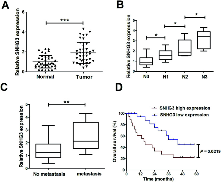
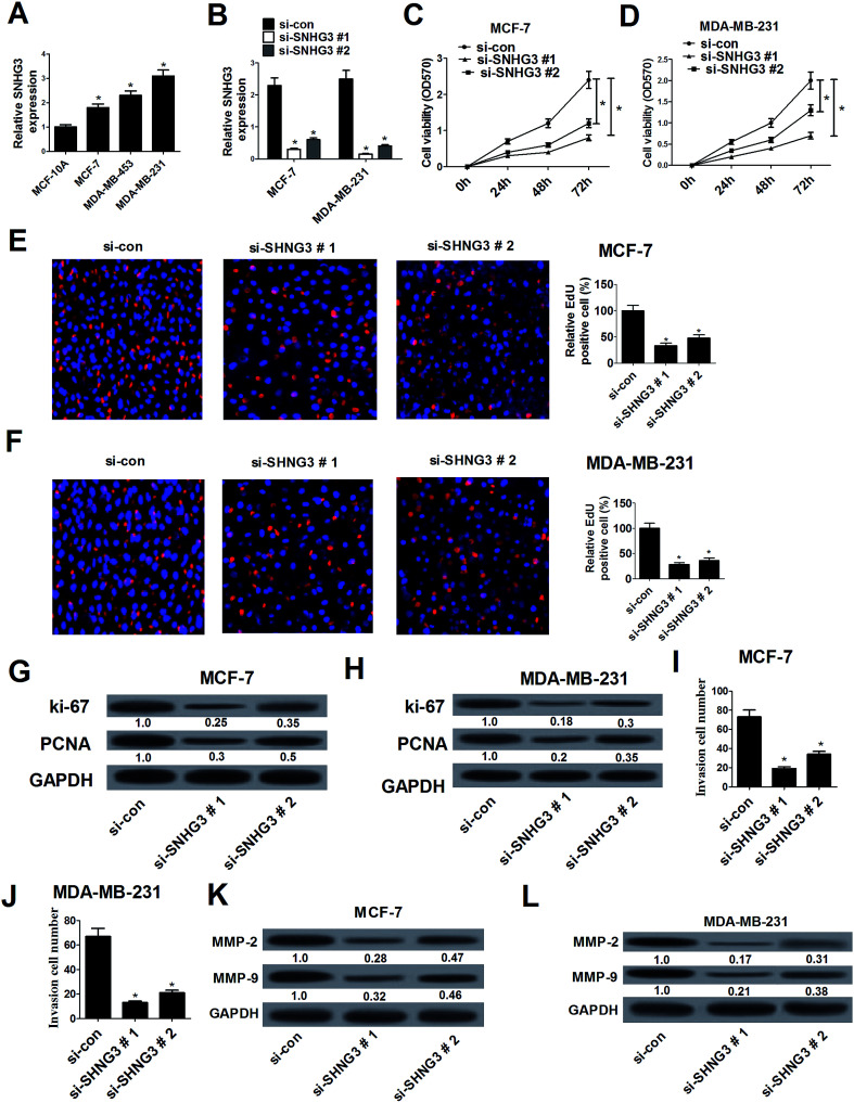
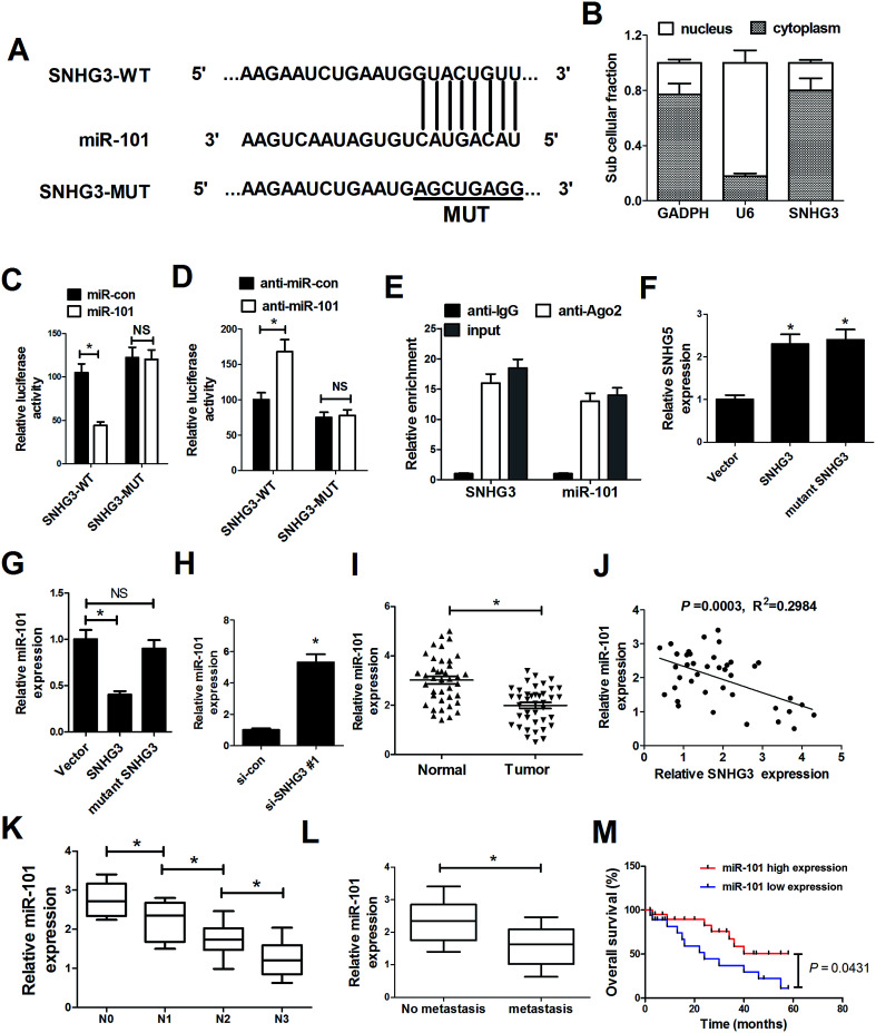
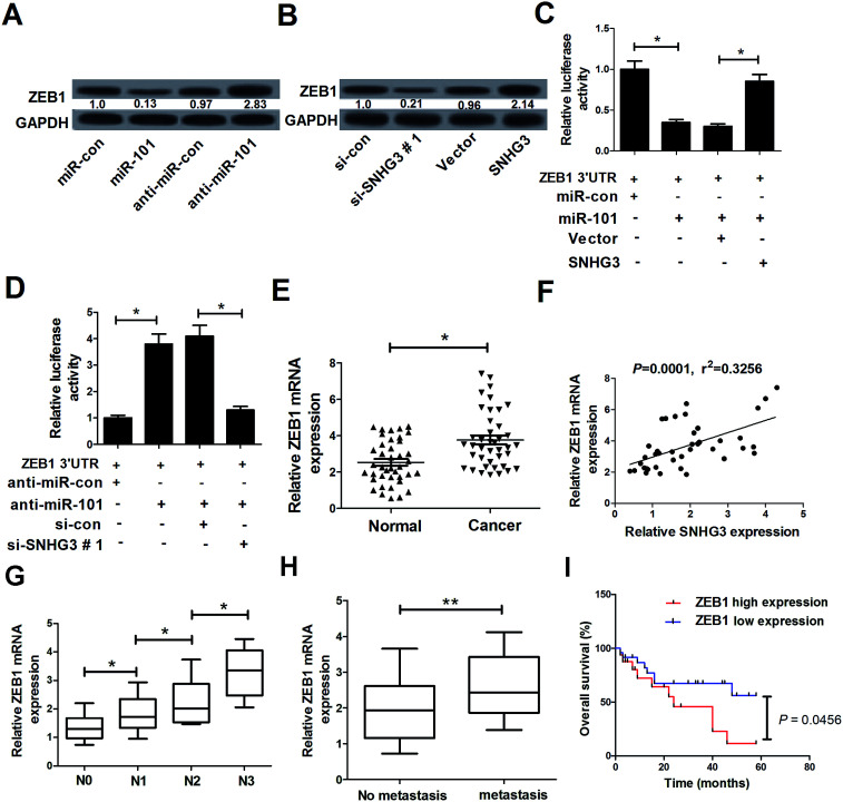
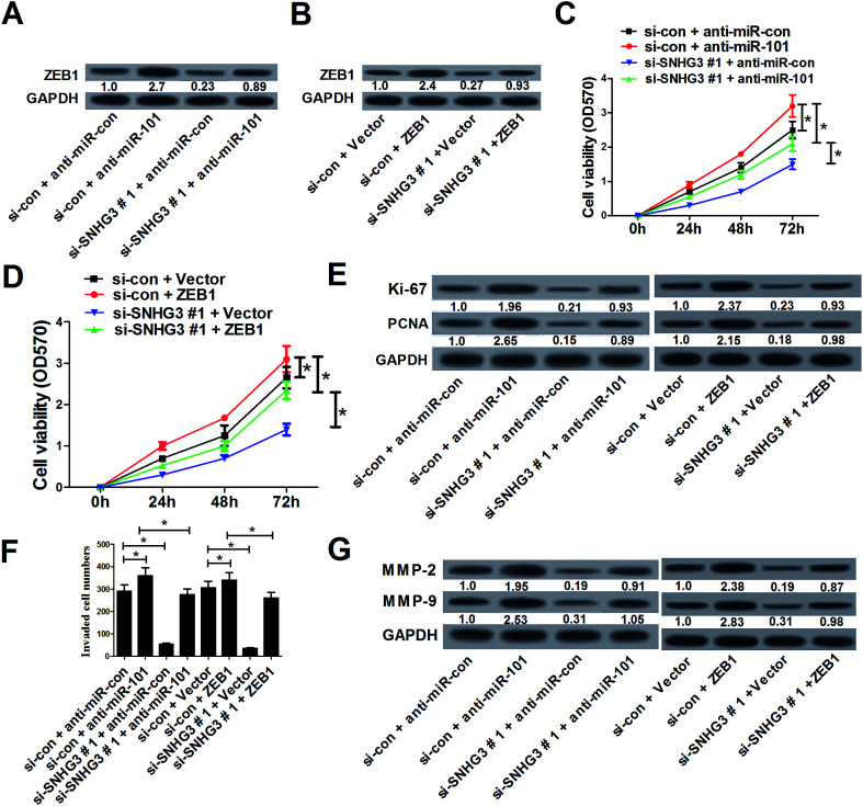
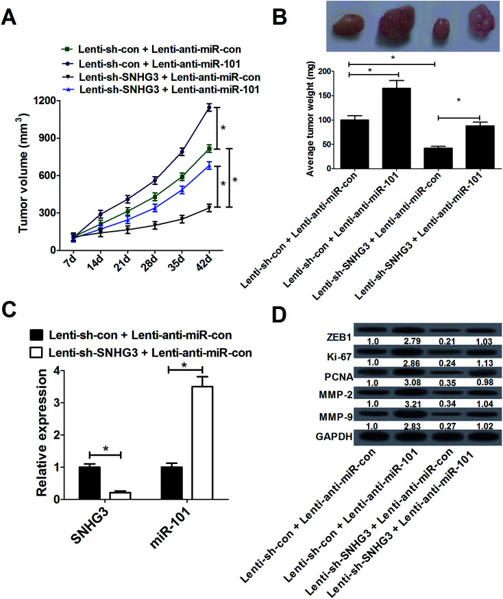
Background: Dysregulated lncRNA expression contributes to the pathogenesis of human tumors via the lncRNAs functioning as oncogenes or tumor suppressors. Small nucleolar RNA host gene 3 (SNHG3) was demonstrated to be upregulated in breast cancer cells. However, the detailed roles and molecular mechanism of SNHG3 in breast cancer are largely unknown. Methods: The expression of SNHG3, miR-101, and zinc finger E-box-binding protein 1 (ZEB1) in breast cancer tissues and cells was detected using qRT-PCR. The effects of SNHG3 on cell proliferation and invasion were evaluated using MTT, EdU, and cell invasion assays. The protein levels of Ki-67, proliferating cell nuclear antigen (PCNA), matrix metalloproteinase MMP-2, and MMP-9 were analyzed using western blot analysis. A luciferase reporter assay and RNA immunoprecipitation (RIP) were performed to explore the interaction between SNHG3, ZEB1 and miR-101. A subcellular fractionation assay was used to detect the subcellular location of SNHG3. Xenograft tumor experiments were conducted to verify the role and mechanism of SNHG3 in breast cancer in vivo. Results: SNHG3 expression was upregulated in breast cancer tissues and correlated with poor prognosis. SNHG3 knockdown suppressed breast cancer cell proliferation and invasion, which was further demonstrated by high levels of proliferation marker proteins Ki-67/PCNA and metastasis-related proteins MMP-2/MMP-9. Additionally, SNHG3 was located in the cytoplasm of breast cancer cells. SNHG3 functioned as a molecular sponge for miR-101 in breast cancer cells. miR-101 was downregulated in breast cancer tissues and negatively correlated with SNHG3 expression. Moreover, ZEB1, a target of miR-101, was positively regulated by SNHG3 in breast cancer cells. ZEB1 mRNA expression was upregulated in breast cancer tissues and positively correlated with SNHG3 expression. Mechanistically, SNHG3 knockdown suppressed cell proliferation and invasion by upregulation of miR-101 and downregulation of ZEB1 expression in breast cancer cells in vitro and in vivo. Conclusion: SNHG3 promoted proliferation and invasion by regulating the miR-101/ZEB1 axis in breast cancer.


Conflict of interest statement

The authors declare that they have no competing interests.

Figures

Fig. 1
Fig. 1. Upregulated SNHG3 was closely correlated with poor prognosis for breast cancer patients. (A) The expression of SNHG3 in 42 paired breast cancer tissues and matched adjacent normal tissues was detected using qRT-PCR. (B) The expression of SNHG3 in different advanced pathological stages N0, N1, N2 and N3. (C) The expression of SNHG3 in 31 breast cancer patients with distant metastasis and 11 breast cancer patients with no distant metastasis. (D) Kaplan–Meier curves and a log-rank test of overall survival for all breast cancer patients. *P < 0.05, **P < 0.01, and ***P < 0.001.
Fig. 2
Fig. 2. SNHG3 silencing suppressed cell proliferation and invasion in breast cancer cells. (A) The expression level of SNHG3 in breast cancer cells (MCF-7, MDA-MB-453 and MDA-MB-231) and the normal breast epithelial cell line MCF-10A was estimated using qRT-PCR. (B) The expression level of SNHG3 in MCF-7 and MDA-MB-231 cells transfected with si-SNHG3#1, si-SNHG3#2 or si-con was evaluated using qRT-PCR. The MTT assay (C and D) and EdU proliferation assay (E and F) were performed to detect cell proliferation at 24 h, 48 h, and 72 h in MCF-7 and MDA-MB-231 cells transfected with si-SNHG3#1, si-SNHG3#2 or si-con. Western blot analysis was conducted to evaluate the protein levels of Ki-67 and PCNA in MCF-7 (G) and MDA-MB-231 (H) cells transfected with si-SNHG3#1, si-SNHG3#2 or si-con. Cell invasion ability was examined using the cell invasion assay in MCF-7 (I) and MDA-MB-231 (J) cells transfected with si-SNHG3#1, si-SNHG3#2 or si-con. The protein levels of MMP-2 and MMP-9 in MCF-7 (K) and MDA-MB-231 (L) cells transfected with si-SNHG3#1, si-SNHG3#2 or si-con were determined using western blot analysis. *P < 0.05.
Fig. 3
Fig. 3. SNHG3 functioned as a molecular sponge for miR-101 in breast cancer cells. (A) The luciferase reporter plasmids containing the predicted wild-type or mutated miR-101 in SNHG3. (B) Subcellular fractionation for SNHG3 in MCF-7 cells, where GAPDH and U6 were used as the controls. (C and D) A luciferase reporter assay was conducted to measure the luciferase activity in MCF-7 cells cotransfected with SNHG3-WT or SNHG3-MUT and miR-101, anti-miR-101, or matched controls. (E) Association between SNHG3 and miR-101 with Ago2 antibody. An anti-Ago2 RIP assay was performed in MCF-7 cell extract and the expressions of SNHG3 and miR-101 were detected using qRT-PCR. qRT-PCR was performed to detect the expressions of SNHG3 (F) and miR-101 (G and H) in MCF-7 cells transfected with SNHG3, mutant SNHG3, Vector, si-SNHG3#1, or si-con. (I) miR-101 expression in 42 paired breast cancer tissues and matched normal tissues. (J) The correlation between SNHG3 and miR-101 expression in breast cancer tissues. (K) The expression of miR-101 in different advanced pathological stages N0, N1, N2 and N3. (L) The expression of miR-101 in 31 breast cancer patients with distant metastasis and 11 breast cancer patients with no distant metastasis. (M) Kaplan–Meier curves and a log-rank test of overall survival for all breast cancer patients. *P < 0.05.
Fig. 4
Fig. 4. The interaction between SNHG3, ZEB1 and miR-101 in breast cancer cells. (A) Western blot analysis of the ZEB1 level in MCF-7 cells transfected with miR-101, anti-miR-101, or matched controls. (B) Western blot analysis of the ZEB1 level in MCF-7 cells introduced to si-SNHG3#1, SNHG3, or the respective controls. (C) MCF-7 cells were cotransfected with ZEB1 3′-UTR and miR-101, miR-con, miR-101 + Vector, or miR-101 + SNHG3. A luciferase reporter assay was performed to detect the luciferase activity at 48 h post-transfection. (D) MCF-7 cells were cotransfected with ZEB1 3′-UTR and anti-miR-101, anti-miR-con, anti-miR-101 + si-con, or anti-miR-101 + si-SNHG3. A luciferase reporter assay was performed to measure the luciferase activity at 48 h post-transfection. (E) ZEB1 mRNA expression in 42 paired breast cancer tissues and adjacent normal tissues. (F) The correlation between SNHG3 and ZEB1 mRNA expression in breast cancer tissues. (G) The expression of ZEB1 in different advanced pathological stages N0, N1, N2 and N3. (H) The expression of ZEB1 in 31 breast cancer patients with distant metastasis and 11 breast cancer patients with no distant metastasis. (I) Kaplan–Meier curves and a log-rank test of overall survival for all breast cancer patients. *P < 0.05, **P < 0.01.
Fig. 5
Fig. 5. SNHG3 knockdown suppressed cell proliferation and invasion by upregulation of miR-101 and downregulation of ZEB1 expression in breast cancer cells in vitro. MCF-7 cells were cotransfected with si-SNHG3#1 or si-con, along with anti-miR-101, anti-miR-con, ZEB1, or Vector, and incubated for 48 h. (A and B) The protein level of ZEB1 in transfected MCF-7 cells. (C and D) Cell proliferation was estimated using an MTT assay in transfected MCF-7 cells. (E) The protein levels of Ki-67 and PCNA in transfected MCF-7 cells. (F) The cell invasion capability was evaluated using a cell invasion assay in transfected MCF-7 cells. (G) The protein levels of MMP-2 and MMP-9 in transfected MCF-7 cells. *P < 0.05.
Fig. 6
Fig. 6. SNHG3 knockdown suppressed breast cancer tumor growth and invasion in vivo by upregulating miR-101 and downregulating ZEB1. Lenti-anti-miR-con or lenti-anti-miR-101 was transduced in lenti-sh-SNHG3 or lenti-sh-con stably transfected MCF-7 cells and then MCF-7 cells were subcutaneously injected into nude mice. (A) Tumor volume was measured every 7 days for 42 days. (B) Forty-two days after injection, the mice were sacrificed and the tumor tissues were isolated and weighed. (C) The expressions of SNHG3 and miR-101 in xenografted tumors. (D) The protein levels of ZEB1, Ki-67, PCNA, MMP-2 and MMP-9 in xenografted tumors were detected using western blot analysis. *P < 0.05.

References

    1. Siegel R. L. Miller K. D. Jemal A. Ca-Cancer J. Clin. 2015;65:5–29. doi: 10.3322/caac.21254. - DOI - PubMed
    1. Kohler B. A. Sherman R. L. Howlader N. Jemal A. Ryerson A. B. Henry K. A. Boscoe F. P. Cronin K. A. Lake A. Noone A. M. Henley S. J. Eheman C. R. Anderson R. N. Penberthy L. J. Natl. Cancer Inst. 2015;107:djv048. doi: 10.1093/jnci/djv048. - DOI - PMC - PubMed
    1. Martin M. Lopez-Tarruella S. Gilarranz Y. J. Breast. 2016;28:161–166. doi: 10.1016/j.breast.2016.05.015. - DOI - PubMed
    1. Alonso D. F. Ripoll G. V. Garona J. Iannucci N. B. Gomez D. E. Curr. Pharm. Biotechnol. 2011;12:1974–1980. - PMC - PubMed
    1. Shi X. Sun M. Liu H. Yao Y. Song Y. Cancer Lett. 2013;339:159–166. doi: 10.1016/j.canlet.2013.06.013. - DOI - PubMed

Publication types