. 2018 Jun 5;8(36):20411-20422.
doi: 10.1039/c8ra00798e. eCollection 2018 May 30.

Promotion of autophagosome-lysosome fusion via salvianolic acid A-mediated SIRT1 up-regulation ameliorates alcoholic liver disease

Affiliations

Promotion of autophagosome-lysosome fusion via salvianolic acid A-mediated SIRT1 up-regulation ameliorates alcoholic liver disease

Xue Shi et al. RSC Adv. .

Abstract

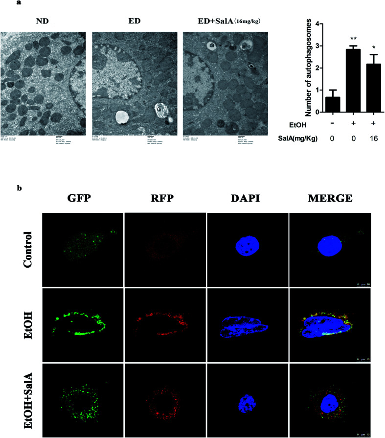
Salvianolic acid A (SalA) is a water-soluble phenolic carboxylic acid extracted from Salvia miltiorrhiza that has extensive pharmacological activities and plays an essential role in liver disease treatment. However, the mechanism of SalA in treating alcoholic liver disease (ALD) remains unclear. Here, we studied the protective effects of SalA on chronic ethanol-induced liver injury involving Sirtuin 1 (SIRT1)-mediated autophagy activation. The results showed that SalA pretreatment reduced the levels of alanine aminotransferase (ALT), aspartate aminotransferase (AST), triglyceride (TG) and cholesterol (TC) in vivo and enhanced hepatic cell viability while mitigating apoptosis and hepatic steatosis in vitro. Furthermore, SalA protected against chronic ethanol-induced liver injury by restoring autophagosome-lysosome fusion, as indicated by the increased expression levels of LC3-II, cathepsin B, lysosomal-associated membrane protein 2 (LAMP-2), and RAB7 and the decreased expression of SQSTM1. More importantly, pretreatment with SalA significantly up-regulated the expression of SIRT1, an NAD+-dependent deacetylase. These increased levels of SIRT1 stimulated autophagy under conditions of chronic ethanol exposure. Interestingly, SIRT1 siRNA abrogated SalA-induced autophagosome-lysosome fusion. This finding indicates that the protective effects of SalA are associated with SIRT1 activation. Collectively, our study demonstrates that SalA pretreatment protects against chronic ethanol-induced liver injury via the SIRT1-mediated restoration of autophagosome-lysosome fusion.


Conflict of interest statement

There are no conflicts to declare.

Figures

Fig. 1
Fig. 1. SalA pretreatment attenuates chronic ethanol-induced liver injury. (a) H&E staining. The experimental groups subjected to H&E staining were as follows: ND; ND + SalA (16 mg kg−1); ED; ED + SalA (8 mg kg−1); and ED + SalA (16 mg kg−1). H&E-stained sections were photographed at 200× magnification. (b) Cell viability. AML-12 cells were pretreated with SalA (0.5, 5 or 50 μM) for 6 h and then treated with ethanol for 24 h. Cell viability was assessed using CCK8 assays. The data are presented as the mean ± S.D. (n ≥ 8). **P < 0.01 versus the control group, ##P < 0.01 versus the ethanol group.
Fig. 2
Fig. 2. SalA mediates autophagy activation. (a) Effects of SalA on AV formation in ALD liver were assessed using the TEM assay (1500×), and autophagosomes were quantified. The data are presented as the mean ± S.D. *P < 0.05 versus the ND group, **P < 0.01 versus the ND group (n = 3). (b) Autophagy flux visualized through confocal microscopy. AML-12 cells were infected with adenoviruses as described in Materials and methods. Autophagy flux was increased when the numbers of both yellow and red puncta were increased in cells, whereas autophagy flux was blocked when the number of only yellow puncta was increased in AML-12 cells.
Fig. 3
Fig. 3. Autophagy activation by SalA exerts a protective effect against chronic ethanol-induced hepatocyte injury. (a and b) SalA-induced autophagy activation decreases lipid accumulation and apoptosis. AML-12 cells were pretreated with SalA (50 μM) or CQ (50 μM) for 6 h before exposure to ethanol (100 mM) for another 24 h. (a) Nile red staining (400×). (b) TUNEL staining (400×). (c) Cell viability. AML-12 cells were pretreated with SalA (50 μM), CQ (50 μM), or EBSS for 6 h and then treated with ethanol for 24 h; afterwards, cell viability was determined. The data are presented as the mean ± S.D. **P < 0.01 versus the control group, ##P < 0.01 versus the ethanol group, &&P < 0.01 versus the ethanol + SalA group (n = 8).
Fig. 4
Fig. 4. SalA promotes autophagy activation to prevent chronic ethanol-induced AV-lysosome fusion damage. (a and b) The expression levels of LC3-II, SQSTM1, the mature form of cathepsin B, RAB7, and LAMP-2 in the liver were evaluated by western blotting. The data are presented as the mean ± S.D. (n = 3). *P < 0.05 versus the ND group, **P < 0.01 versus the ND group, #P < 0.05 versus the ED group, ##P < 0.01 versus the ED group.
Fig. 5
Fig. 5. SIRT1 mediates the restoration of AV-lysosome fusion by SalA in ethanol-treated AML-12 cells. (a) AML-12 cells were transfected with SIRT1-specific siRNA or control siRNA for 48 h before treatment with SalA (50 μM). Then, the transfected cells were exposed to ethanol (100 mM) for another 24 h. SIRT1, LAMP-2, and RAB7 protein levels in cellular lysates were evaluated by western blotting. The data are presented as the mean ± S.D. **P < 0.01 versus the si-control + ethanol group (n = 3). (b) Autophagy flux visualized using confocal microscopy. AML-12 cells were infected with adenoviruses as described in Materials and methods.

Similar articles

Cited by

References

    1. Louvet A. Mathurin P. Alcoholic liver disease: mechanisms of injury and targeted treatment. Nat. Rev. Gastroenterol. Hepatol. 2015;12:231–242. doi: 10.1038/nrgastro.2015.35. - DOI - PubMed
    1. Vuittonet C. L. Halse M. Leggio L. Fricchione S. B. Brickley M. Haass-Koffler C. L. Tavares T. Swift R. M. Kenna G. A. Pharmacotherapy for alcoholic patients with alcoholic liver disease. Am. J. Health-Syst. Pharm. 2014;71:1265–1276. doi: 10.2146/ajhp140028. - DOI - PMC - PubMed
    1. Bruha R. Dvorak K. Petrtyl J. Alcoholic liver disease. World J Hepatol. 2012;4:81–90. doi: 10.4254/wjh.v4.i3.81. - DOI - PMC - PubMed
    1. O'Shea R. S. Dasarathy S. McCullough A. J. Practice Guideline Committee of the American Association for the Study of Liver and Practice Parameters Committee of the American College of, Alcoholic liver disease. Hepatology. 2010;51:307–328. doi: 10.1002/hep.23258. - DOI - PubMed
    1. Bailey S. M. Cunningham C. C. Contribution of mitochondria to oxidative stress associated with alcoholic liver disease. Free Radical Biol. Med. 2002;32:11–16. doi: 10.1016/S0891-5849(01)00769-9. - DOI - PubMed