. 2018 Jun 13;8(39):21889-21904.
doi: 10.1039/c8ra02735h.

Osteoinductive 3D scaffolds prepared by blend centrifugal spinning for long-term delivery of osteogenic supplements

Affiliations

Osteoinductive 3D scaffolds prepared by blend centrifugal spinning for long-term delivery of osteogenic supplements

Lukasova Vera et al. RSC Adv. .

Abstract

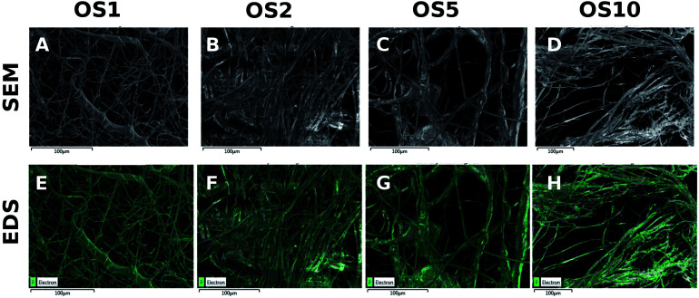
Bone regeneration is a long-term process requiring proper scaffolding and drug delivery systems. The current study delivers a three-dimensional (3D) scaffold prepared by blend centrifugal spinning loaded with the osteogenic supplements (OS) β-glycerol phosphate, ascorbate-2-phosphate and dexamethasone. The OS were successfully encapsulated into a fibrous scaffold and showed sustained release for 30 days. Furthermore, biological testing showed the osteoinductive properties of the scaffolds on a model of human mesenchymal stem cells and stimulatory effect on a model of osteoblasts. The osteoinductive properties were further proved in vivo in critical size defects of rabbits. The amount of bone trabecules was bigger compared to control fibers without OS. The results indicate that due to its long-term drug releasing properties, single step fabrication process and 3D structure, the system shows ideal properties for use as a cell-free bone implant in tissue-engineering.


Conflict of interest statement

There are no conflicts to declare.

Figures

Fig. 1
Fig. 1. Images from scanning electron microscopy of scaffolds. Micrograph of scaffold morphology with lower magnification (scale bar 20 μm) (A–E), detailed micrograph of fibers showing phosphate microdomains (scale bar 5 μm) (F–J), detailed fiber surface after 31 days washing in PBS (scale bar 5 μm) (K–O).
Fig. 2
Fig. 2. Spectra of prepared scaffolds measured by FTIR-ATR spectroscopy.
Fig. 3
Fig. 3. HR-SEM analysis of PCL scaffolds. Images from scanning electron microscopy (A–D). Distribution of phosphate in PCL scaffolds with encapsulated OS, measured by HR-SEM with EDS detector (E–H).
Fig. 4
Fig. 4. Release of phosphate from fibers. Absolute release of phosphate over time (A). Cumulative release of phosphate from fibers (B).
Fig. 5
Fig. 5. Saos2 adhesion, proliferation, metabolic activity and type I collagen expression on scaffolds. Cell adhesion and proliferation was measured using quantification of DNA (A). Metabolic activity was determined from MTS assay (B). Relative expression of type I collagen was analyzed by qPCR (C). Statistical significance is shown above the columns (*p < 0.05; **p < 0.01).
Fig. 6
Fig. 6. Immunofluorescence staining of osteocalcin. Saos2 stained on day 21 (A–F) and hMSCs stained on day 14 (G–L). Osteocalcin was stained using indirect fluorescence (green color) and cell nuclei using propidium iodide (red color) and visualized using a confocal microscope. Magnification 200×, scale 100 μm.
Fig. 7
Fig. 7. hMSCs adhesion, proliferation and metabolic activity on scaffolds. Cell adhesion and proliferation was measured using quantification of DNA (A). Metabolic activity was determined from MTS assay (B). Statistical significance is shown above the columns (*p < 0.05; **p < 0.01).
Fig. 8
Fig. 8. Visualization of hMSCs adhesion and distribution on scaffolds using a confocal microscope. Cell nuclei were stained using propidium iodide (red color) and cell internal membranes using DiOC3 (green color). Magnification 200×, scale 100 μm (A–L). hMSCs adhesion and interaction on fibrous scaffolds visualized using scanning electron microscopy. Magnification 3000×, scale 20 μm (M–X).
Fig. 9
Fig. 9. Osteogenic differentiation of hMSCs was detected using ALP activity measurement and qPCR analysis of osteogenic markers. ALP activity of hMSCs (A), relative expression of RunX2 (B), type I collagen (C) and osteocalcin (D) was determined by qPCR analysis. Statistical significance is shown above the columns (*p < 0.05; **p < 0.01).
Fig. 10
Fig. 10. Between-group comparison of bone volume. Bone quantity was expressed as volume fraction (Vv) of bone tissue within the whole reference volume of the defect (Box and Whisker plot showing mean, standard error, and standard deviation. The differences were considered statistically significant as *p < 0.05). Bone tissue and collagen connective tissue contain type I collagen were stained using picrosirius red (A). In group I (B) and II (C), newly formed bone trabecules were found in outer compartment of bone defect. In group III (D), there was mainly adipose tissue and small vessels in bone defect. Newly formed bone trabecules were stained using Verhoeff's haematoxylin with green trichrome. In group I (E) and II (F), a few newly formed bone trabecules (red arrows) were found in outer compartment of bone defect. In group III (G), there was mainly adipose tissue in bone defect. Scale 1000 μm.

References

    1. Lucas R. Martins A. Severo M. Silva P. Monjardino T. Gaio A. R. Cooper C. Barros H. Sci. Rep. 2017;7:3754. doi: 10.1038/s41598-017-03847-x. - DOI - PMC - PubMed
    1. Mehta M. Schmidt-Bleek K. Duda G. N. Mooney D. J. Adv. Drug Delivery Rev. 2012;64:1257–1276. doi: 10.1016/j.addr.2012.05.006. - DOI - PMC - PubMed
    1. Wiltfang J. Kloss F. R. Kessler P. Nkenke E. Schultze-Mosgau S. Zimmermann R. Schlegel K. A. Clin. Oral Implants Res. 2004;15:187–193. doi: 10.1111/j.1600-0501.2004.00980.x. - DOI - PubMed
    1. Banwart J. C. Asher M. A. Hassanein R. S. Spine. 1995;20:1055–1060. doi: 10.1097/00007632-199505000-00012. - DOI - PubMed
    1. Dimitriou R. Mataliotakis G. I. Angoules A. G. Kanakaris N. K. Giannoudis P. V. Injury. 2011;42(suppl 2):S3–S15. doi: 10.1016/j.injury.2011.06.015. - DOI - PubMed