. 2018 Mar 6;8(17):9394-9401.
doi: 10.1039/c8ra00232k. eCollection 2018 Feb 28.

Tailor-made spider-eggcase-silk spheres for efficient lysosomal drug delivery

Affiliations

Tailor-made spider-eggcase-silk spheres for efficient lysosomal drug delivery

Jianming Chen et al. RSC Adv. .

Abstract

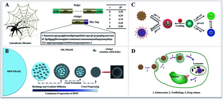
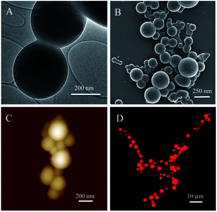
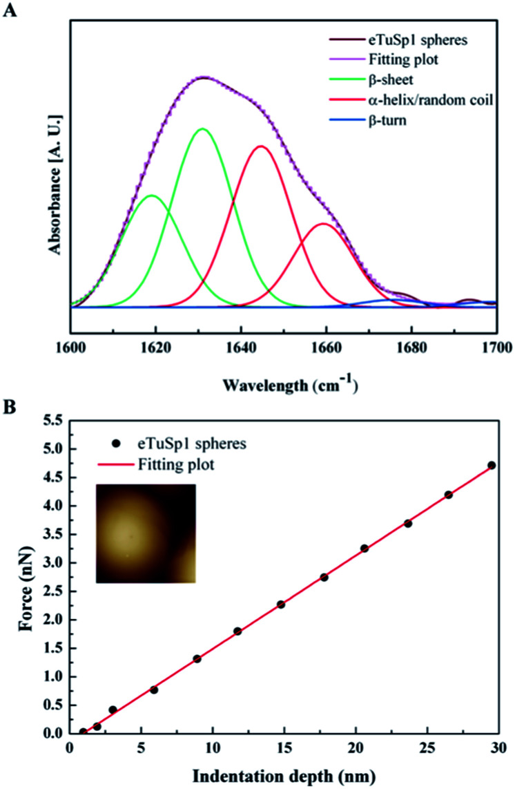
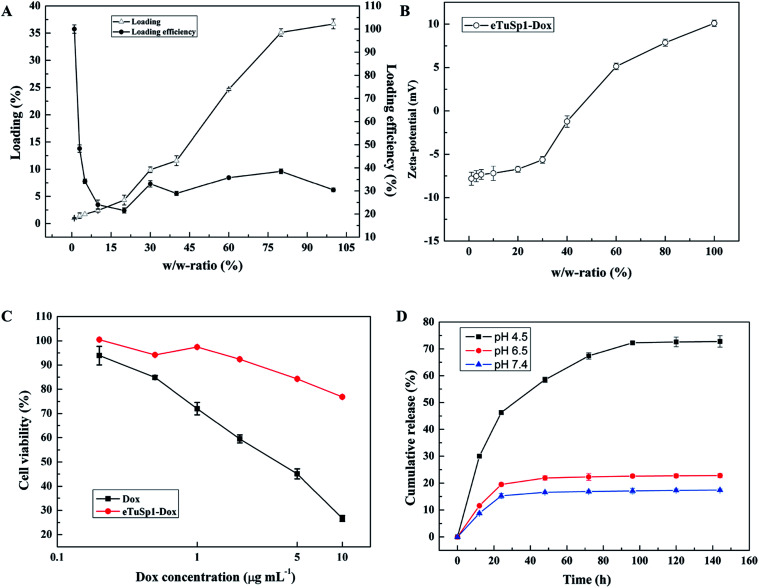
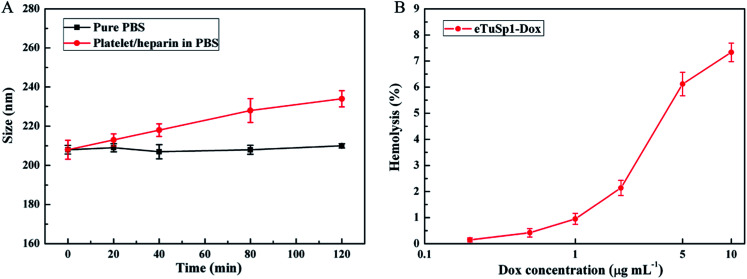
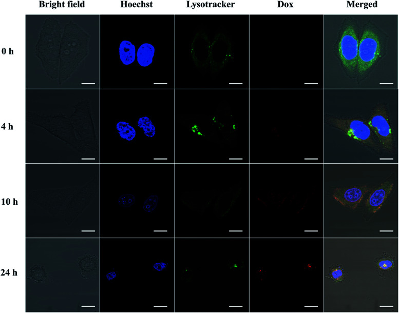
Spider silks are attractive biopolymers due to their excellent mechanical properties and biomimetic potential. To optimize the electrostatic interaction for lysosomal drug delivery, a spider-eggcase-silk protein was genetically engineered using 5× His Tag with a tailor-made isoelectric point of 4.8. By a facile HFIP-on-oil method, silk spheres were assembled as rapidly as 10 s. After the post-treatment of ethanol, silk spheres were determined with an improved compressive modulus by AFM indentation. Under incubation of silk spheres in a Doxorubicin solution, a maximum of 35% loading and average of 30% loading efficiency were determined. In the cytotoxicity experiment, silk spheres exhibited intrinsic biocompatibility and showed good control of the loaded drug in the neutral PBS solution. Significantly, by 96 h, the accumulative drug release at pH 4.5 was approximately 4.5-fold higher than that at pH 7.4. By conducting the platelet adhesion and hemolysis assay, Doxorubicin-loaded silk spheres exhibited good hemocompatibility. To further demonstrate this release behavior, within 24 h, Doxorubicin-loaded silk spheres were efficiently delivered to lysosomes and then released the payload to the nuclei of Hela cells.


Conflict of interest statement

There are no conflicts to declare.

Figures

Fig. 1
Fig. 1. Schematic illustration of the design of eTuSp1 and the application of eTuSp1 spheres in lysosomal delivery systems. (A) Engineered spider-eggcase-silk proteins from a black widow spider. In comparison to bare proteins, the isoelectric point of recombinant proteins can be flexibly regulated from 3.35 to 5.56 by fusing with different numbers of His Tags. (B) Fabrication process of eTuSp1 spheres by the HFIP-on-oil method. (C) The pI-mediated drug loading and release of eTuSp1-Dox spheres. Silk spheres are loaded with Dox (red) in the negative (green) state and release the drug in the positive (purple) state. (D) Internalization and intracellular drug release. (1) Endocytosis; (2) trafficking; (3) drug release.
Fig. 2
Fig. 2. The morphology structure of silk spheres and their potential in drug loading. (A) TEM image of compact eTuSp1 spheres. (B) FE-SEM image of dried eTuSp1 spheres. (C) AFM image of eTuSp1 spheres. (D) CLSM image of eTuSp1-Dox spheres.
Fig. 3
Fig. 3. The secondary structure and compressive modulus of eTuSp1 spheres. (A) ATR-FTIR spectra of eTuSp1 spheres. (B) Mechanical property of eTuSp1 spheres. The compressive force is investigated as a function of indentation depth. The inset shows the AFM image recorded for the local force measurement on a single sphere.
Fig. 4
Fig. 4. Loading and release behaviors of eTuSp1 spheres. (A) Loading and loading efficiency of eTuSp1 spheres. (B) Zeta-potential of eTuSp1-Dox spheres with regard to different w/w-ratios. (C) Cytotoxicity of free Dox and eTuSp1-Dox spheres. The Dox concentration of eTuSp1-Dox spheres is equivalent to that of the free Dox. (D) Cumulative release of eTuSp1-Dox spheres at different pH media.
Fig. 5
Fig. 5. Hemocompatibility of eTuSp1-Dox spheres. (A) platelet adhesion assay. The size of eTuSp1-Dox spheres is evaluated when exposed to pure PBS and platelet/heparin solution. (B) Hemolysis assay. The hemolysis of eTuSp1-Dox spheres is conducted with Dox concentrations ranging from 0.2 to 10 μg mL−1.
Fig. 6
Fig. 6. Lysosomal delivery of eTuSp1-Dox spheres in Hela cells. Hoechst and Lysotracker are used to strain the cell nucleus (blue) and lysosomes (green), respectively. The trafficking of the Dox in the intracellular environment is recorded in 24 h by the aid of fluorescence detection. The scale bar represents 10 μm.

Similar articles

Cited by

References

    1. Duncan R. Nat. Rev. Cancer. 2006;6:688–701. doi: 10.1038/nrc1958. - DOI - PubMed
    1. Berkland C. Kim K. K. Pack D. W. J. Controlled Release. 2001;73:59–74. doi: 10.1016/S0168-3659(01)00289-9. - DOI - PubMed
    1. Herrmann J. Bodmeier R. J. Controlled Release. 1995;36:63–71. doi: 10.1016/0168-3659(95)00051-9. - DOI
    1. Ulbrich K. Šubr V. r. Adv. Drug Delivery Rev. 2004;56:1023–1050. doi: 10.1016/j.addr.2003.10.040. - DOI - PubMed
    1. Gillies E. R. Jonsson T. B. Fréchet J. M. J. Am. Chem. Soc. 2004;126:11936–11943. doi: 10.1021/ja0463738. - DOI - PubMed