. 2018 Feb 20;8(15):8164-8172.
doi: 10.1039/c7ra12815k. eCollection 2018 Feb 19.

MiR-29c inhibits HCV replication via activation of type I IFN response by targeting STAT3 in JFH-1-infected Huh7 cells

Affiliations

MiR-29c inhibits HCV replication via activation of type I IFN response by targeting STAT3 in JFH-1-infected Huh7 cells

Yanjing Wang et al. RSC Adv. .

Abstract

Background: MiR-29c, a member of the miR-29 family, has been recognized to play an important role in hepatitis C virus (HCV) infection. However, the underlying molecular mechanism of miR-29c involved in HCV replication is not fully understood. Methods: RT-qPCR assay was used to detect the expression pattern of miR-29c and signal transducer and activator of transcription 3 (STAT3) mRNA in JFH-1-infected Huh7 cells. HCV replication was evaluated by the expression of HCV RNA, non-structural protein 5A (NS5A) and non-structural protein 3 (NS3). Dual-Luciferase Reporter assay was applied to search for the candidate target mRNAs of miR-29c. Western blot assay was performed to detect the protein level of double-stranded RNA-dependent protein kinase R (PKR), (2'-5')-oligoadenylate synthetase (OAS) and interferon regulatory transcription factor 1 (IRF1). Results: miR-29c expression was down-regulated, and STAT3 mRNA and protein expressions were up-regulated in JFH-1-infected Huh7 cells. MiR-29c overexpression or STAT3 knockdown repressed HCV replication, while miR-29c depletion or STAT3 upregulation promoted HCV replication. Additionally, STAT3 was a direct target of miR-29c, and miR-29c suppressed STAT3 protein expression in Huh7 cells. Moreover, STAT3 overexpression reversed miR-29c-mediated suppression on HCV replication. Furthermore, the anti-miR-29c-mediated inhibitory effect on type I IFN response was abated following STAT3 knockdown. Conclusions: miR-29c might repress HCV infection via promoting type I IFN response by targeting STAT3 in JFH-1-infected Huh7 cells, offering a promising avenue for HCV treatment.


Conflict of interest statement

The authors have no conflict of interest to declare.

Figures

Fig. 1
Fig. 1. Overexpression of miR-29c repressed HCV replication in JFH-1-infected Huh7 cells. (A) RT-qPCR assay was performed to detect the expression pattern of miR-29c in JFH-1-infected Huh7 cells. (B) The expression level of miR-29c was detected in Huh7 cells transfected with miR-29c mimics or anti-miR-29c. (C and D) Huh7 cells transfected with miR-29c mimics, anti-miR-29c or respective control were infected with JFH-1 (0.01 MOI) for 24 h, followed by the detection of HCV RNA expression by RT-qPCR (C), NS3 and NS5A proteins expression by western blot assay (D). GAPDH was used as the internal reference. *P < 0.05 vs. respective control.
Fig. 2
Fig. 2. STAT3 was a direct target of miR-29c. (A) The wild and mutant 3′-UTR of STAT3 mRNA with the putative binding sites of miR-29c. (B and C) Dual-Luciferase Reporter assay was performed to explore whether STAT3 was a direct target of miR-29c. Wild-type or mutant-type pGL3-STAT3 luciferase vectors (WT-STAT3 or MUT-STAT3) were constructed and transfected into Huh7 cells together with miR-con or miR-29c mimics (B), anti-miR-con or anti-miR-29c (C). (D) Huh7 cells were transfected with miR-con, miR-29c mimics, anti-miR-con or anti-miR-29c for 48 h, followed by the measurement of STAT3 protein expression. *P < 0.05 vs. corresponding control.
Fig. 3
Fig. 3. STAT3 overexpression promoted HCV replication in JFH-1-infected Huh7 cells. RT-qPCR assay of STAT3 mRNA expression (A) and western blot assay of STAT3 protein expression (B) in JFH-1-infected Huh7 cells. (C–E) At 48 h after transfection with pDNA-STAT3, Huh7 cells were infected with JFH-1 (0.01 MOI) for 24 h, followed by the evaluation of STAT3 protein expression (C), HCV RNA expression (D), NS3 and NS5A protein expressions (E). (F–H) Huh7 cells were pretreated with 0, 10, and 20 μM of LIF for 1 h before infection with JFH-1 (0.01 MOI) for 24 h, followed by the assessment of STAT3-Y705 phosphorylation (F), HCV RNA expression (G), NS3 and NS5A proteins expression (H). *P < 0.05 vs. corresponding control.
Fig. 4
Fig. 4. STAT3 silence repressed HCV replication in JFH-1-infected Huh7 cells. RT-qPCR assay of STAT3 mRNA expression (A) and western blot assay of STAT3 expression (B) in Huh7 cells transfected with si-STAT3 or si-con. (C and D) Huh7 cells transfected with si-STAT3 or si-con were infected with JFH-1 for 24 h, followed by the analysis of HCV RNA expression (C), NS5A and NS3 protein expressions (D). (E–G) Huh7 cells were pretreated with various concentrations (0, 10, and 20 μM) of AG490 for 1 h and then infected with JFH-1 for 24 h, followed by the detection of STAT3-Y705 phosphorylation (E), HCV RNA expression (F), NS3 and NS5A proteins expression (G). *P < 0.05 vs. respective control.
Fig. 5
Fig. 5. Restoration of STAT3 expression reversed miR-29c-mediated inhibitory effect on HCV replication in JFH-1-infected Huh7 cells. (A and B) Huh7 cells transfected with miR-29c mimics alone, or together with STAT3 were infected with JFH-1 (0.01 MOI) for 24 h, followed by the assay of HCV RNA expression (A), NS3 and NS5A proteins expression (B). (C and D) Huh7 cells were transfected with anti-miR-29c alone, or together with si-STAT3 prior to infection with JFH-1 (0.01 MOI) for 24 h, followed by the determination of HCV RNA expression (C), NS3 and NS5A proteins expression (D). *P < 0.05 vs. respective control.
Fig. 6
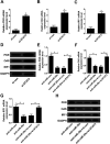
Fig. 6. Anti-miR-29c-mediated suppression on type I IFN response was abated following STAT3 knockdown. RT-qPCR assay of PKR mRNA (A), OAS mRNA (B), IRF1 mRNA (C) expression and western bolt assay of PKR/OAS/IRF1 proteins expression (D) in Huh7 cells transfected with si-STAT3 or si-con. RT-qPCR assay of PKR mRNA (E), OAS mRNA (F), IRF1 mRNA (G) expression and western bolt assay of PKR/OAS/IRF1 proteins expression (H) in Huh7 cells transfected with anti-miR-29c alone, or together with si-STAT3. *P < 0.05 vs. corresponding control.

Similar articles

Cited by

References

    1. Wang S. H., Yeh S. H. and Chen P. J., Hepatitis C Virus II, 2016, pp. 109–136
    1. Wakita T. Methods Mol. Biol. 2009;510:305–327. - PubMed
    1. Welzel T. Petersen J. Herzer K. Ferenci P. Gschwantler M. Cornberg M. Ingiliz P. Berg T. Spengler U. Weiland O. J. Hepatol. 2016;64:S781–S782.
    1. Horsleysilva J. L. Vargas H. E. Gastroenterol. Hepatol. 2017;48:22–31. - PubMed
    1. Maan R. van der Meer A. J. F1000Research. 2016;5:367. doi: 10.12688/f1000research.7399.1. - DOI - PMC - PubMed