

Hematite iron oxide nanoparticles: apoptosis of myoblast cancer cells and their arithmetical assessment
- PMID: 35542163
- PMCID: PMC9082308
- DOI: 10.1039/c8ra02613k
Hematite iron oxide nanoparticles: apoptosis of myoblast cancer cells and their arithmetical assessment
Abstract
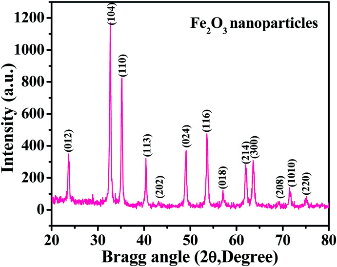
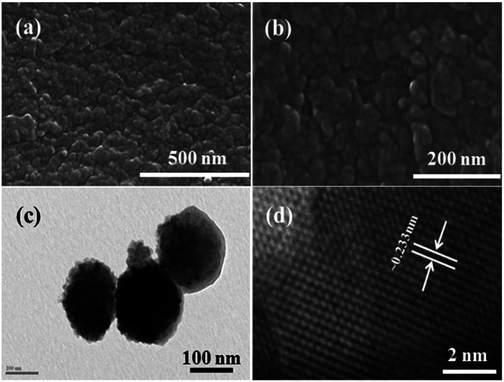
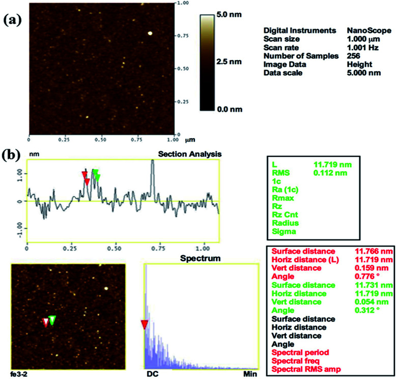
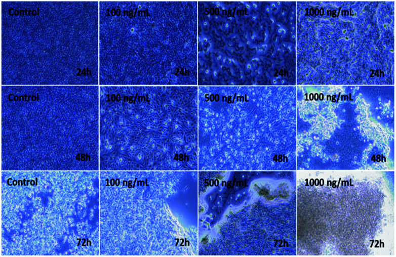
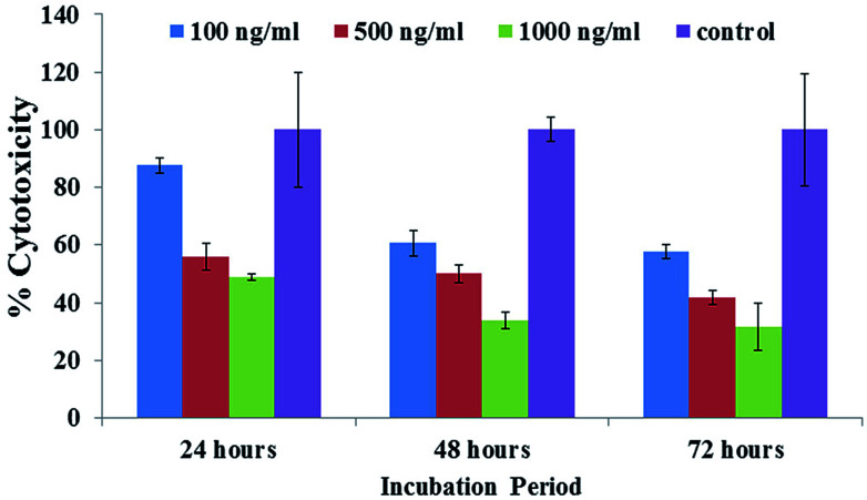
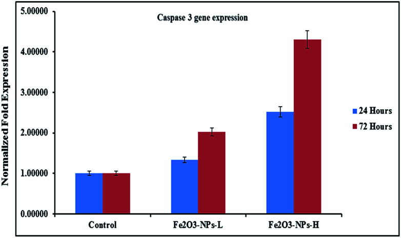
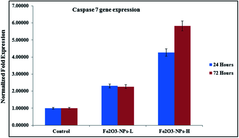
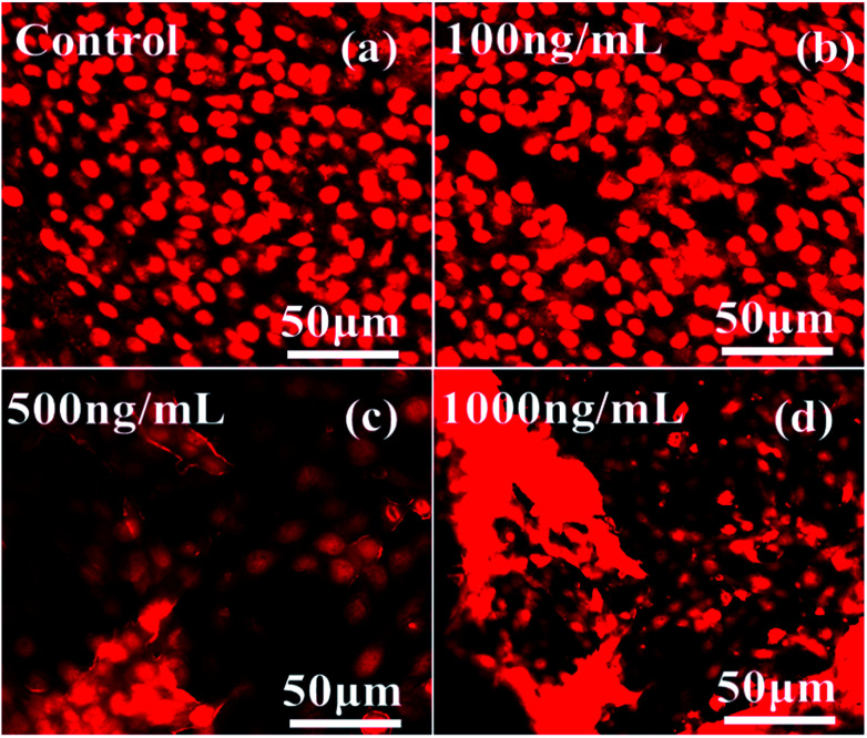
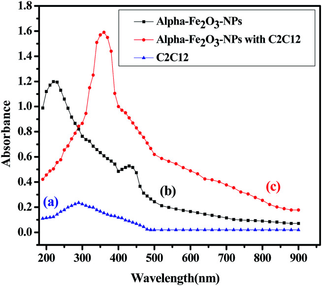
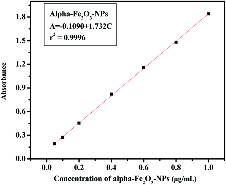
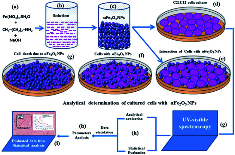
Hematite (α-Fe2O3) forms iron oxide nanoparticles (NPs) which are thermally stable and have various electrochemical and optochemical applications. Due to their wide applicability, the present work was designed to form the hematite phase of iron oxide (αFe2O3NPs) NPs prepared via a solution process. Their cytological performance was checked with C2C12 cells. The crystalline property of the NPs was examined with X-ray diffraction patterns (XRD) and it was found that the size of the particles formed ranged from 12 to 15 nm. Structural information was also identified via field emission scanning electron microscopy (FESEM) and transmission electron microscopy (TEM), which again confirmed that the size of each NP is about 12-15 nm. Surface topographical analysis was done via atomic force microscopy (AFM), which reveals that the size of the distance between two particles is in the range of 12 ± 3 nm. The C2C12 cells were cultured in a humidified environment with 5% CO2 and were checked via a microscope. The αFe2O3NPs were used for cytotoxic evaluation against C2C12 cells. A MTT (3-(4,5-dimethyl thiazol-2-yl)-2,5-diphenyltetrazolium bromide) assay was utilized to check the viability of cells in a dose-dependent (100 ng mL-1, 500 ng mL-1 or 1000 ng mL-1) manner. The morphology of the cells under the influence of αFe2O3NPs for live and dead cells in a wet environment was confirmed via confocal laser scanning microscopy (CLSM). The apoptosis caused due to the αFe2O3NPs was evaluated in presence of caspases 3/7 with GAPDH genes, which confirmed the upregulation that is responsible in caspase 3/7 genes, with treatment of C2C12 at low (500 ng mL-1) and high (1000 ng mL-1) doses of αFe2O3NPs. Analytical studies were also performed to authenticate the obtained data for αFe2O3NPs using parameters such as precision, accuracy, linearity, limits of detection (LOD) and limit of quantitation (LOQ), quantitative recoveries and relative standard deviation (RSD). The analyses play a significant role in investigating the large effect of αFe2O3NPs on C2C12 cells.
This journal is © The Royal Society of Chemistry.
Conflict of interest statement
The authors declare that there are no conflicts of interest.
Figures
Similar articles
-
Statistical analysis of gold nanoparticle-induced oxidative stress and apoptosis in myoblast (C2C12) cells.Colloids Surf B Biointerfaces. 2014 Nov 1;123:664-72. doi: 10.1016/j.colsurfb.2014.10.012. Epub 2014 Oct 12. Colloids Surf B Biointerfaces. 2014. PMID: 25456994
-
Microwave plasma-assisted silicon nanoparticles: cytotoxic, molecular, and numerical responses against cancer cells.RSC Adv. 2019 Apr 30;9(23):13336-13347. doi: 10.1039/c8ra10185j. eCollection 2019 Apr 25. RSC Adv. 2019. PMID: 35520784 Free PMC article.
-
Apoptosis induced by copper oxide quantum dots in cultured C2C12 cells via caspase 3 and caspase 7: a study on cytotoxicity assessment.Appl Microbiol Biotechnol. 2013 Jun;97(12):5545-53. doi: 10.1007/s00253-013-4724-1. Epub 2013 Mar 7. Appl Microbiol Biotechnol. 2013. PMID: 23467821
-
One-Pot Facile Synthesis of Pt Nanoparticles Using Cultural Filtrate of Microgravity Simulated Grown P. chrysogenum and Their Activity on Bacteria and Cancer Cells.J Nanosci Nanotechnol. 2018 May 1;18(5):3110-3125. doi: 10.1166/jnn.2018.14661. J Nanosci Nanotechnol. 2018. PMID: 29442810
-
Magnetic materials as sorbents for metal/metalloid preconcentration and/or separation. A review.Anal Chim Acta. 2013 Jul 30;789:1-16. doi: 10.1016/j.aca.2013.04.021. Epub 2013 Apr 29. Anal Chim Acta. 2013. PMID: 23856225 Review.
Cited by
-
The Use of Deep Eutectic Solvents for the Synthesis of Iron Oxides Nanoparticles: A Driving Force for Materials Properties.Chemistry. 2025 May;31(25):e202500089. doi: 10.1002/chem.202500089. Epub 2025 Mar 31. Chemistry. 2025. PMID: 40105897 Free PMC article.
-
Effective degradation of tetracycline in aqueous solution by an electro-Fenton process using chemically modified carbon/α-FeOOH as catalyst.J Environ Health Sci Eng. 2024 May 18;22(1):313-327. doi: 10.1007/s40201-024-00902-4. eCollection 2024 Jun. J Environ Health Sci Eng. 2024. PMID: 38887776 Free PMC article.
-
Development of Magnetic Sponges Using Steel Melting on 3D Carbonized Spongin Scaffolds Under Extreme Biomimetics Conditions.Biomimetics (Basel). 2025 May 28;10(6):350. doi: 10.3390/biomimetics10060350. Biomimetics (Basel). 2025. PMID: 40558319 Free PMC article.
-
Biogenic iron oxide nanoparticles enhance callogenesis and regeneration pattern of recalcitrant Cicer arietinum L.PLoS One. 2020 Dec 1;15(12):e0242829. doi: 10.1371/journal.pone.0242829. eCollection 2020. PLoS One. 2020. PMID: 33259506 Free PMC article.
-
Role of Solvents in Iron Nanoparticle Synthesis: Analyzing Water and 1‑Methyl-2-Pyrrolidone with Green Tea Extract as a Reducing Agent.ACS Omega. 2025 Jul 10;10(28):30985-30995. doi: 10.1021/acsomega.5c03914. eCollection 2025 Jul 22. ACS Omega. 2025. PMID: 40727754 Free PMC article.
References
-
- Atyam K. K. Ghosh A. Mukherjee K. Majumder S. B. Hematite iron oxide nano-particles: facile synthesis and their chemi-resistive response towards hydrogen. Mater. Res. Express. 2015;2:055901. doi: 10.1088/2053-1591/2/5/055901. - DOI
-
- Kim K. Kim I. H. Yoon K. Y. Lee J. Jang J. H. α-Fe2O3 on patterned fluorine doped tin oxide for efficient Photo electrochemical water splitting. J. Mater. Chem. A. 2015;3:7706–7709. doi: 10.1039/C5TA00027K. - DOI
LinkOut - more resources
Full Text Sources
Other Literature Sources
Research Materials
Miscellaneous