. 2019 Nov 13;9(64):37127-37135.
doi: 10.1039/c9ra08245j.

A Maitake (Grifola frondosa) polysaccharide ameliorates Alzheimer's disease-like pathology and cognitive impairments by enhancing microglial amyloid-β clearance

Affiliations

A Maitake (Grifola frondosa) polysaccharide ameliorates Alzheimer's disease-like pathology and cognitive impairments by enhancing microglial amyloid-β clearance

Yao Bai et al. RSC Adv. .

Abstract

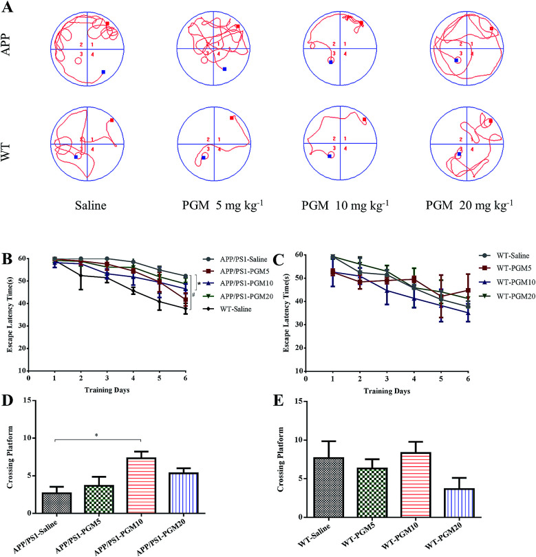
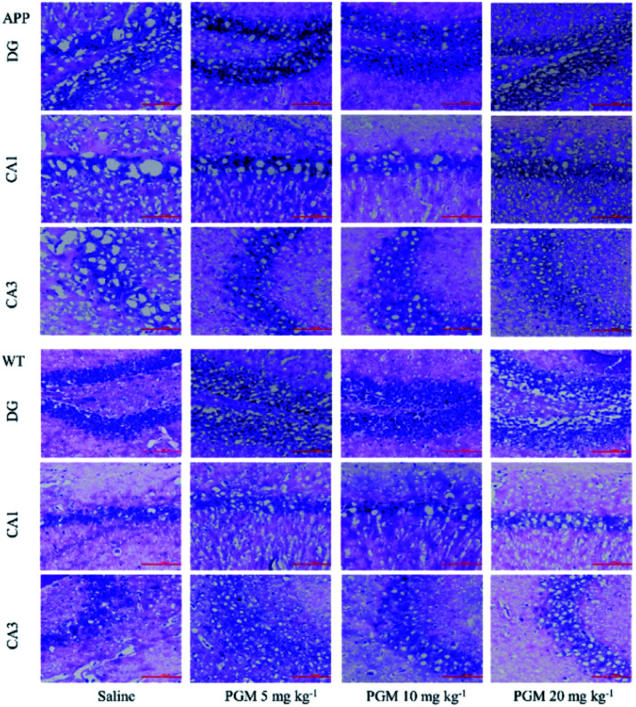
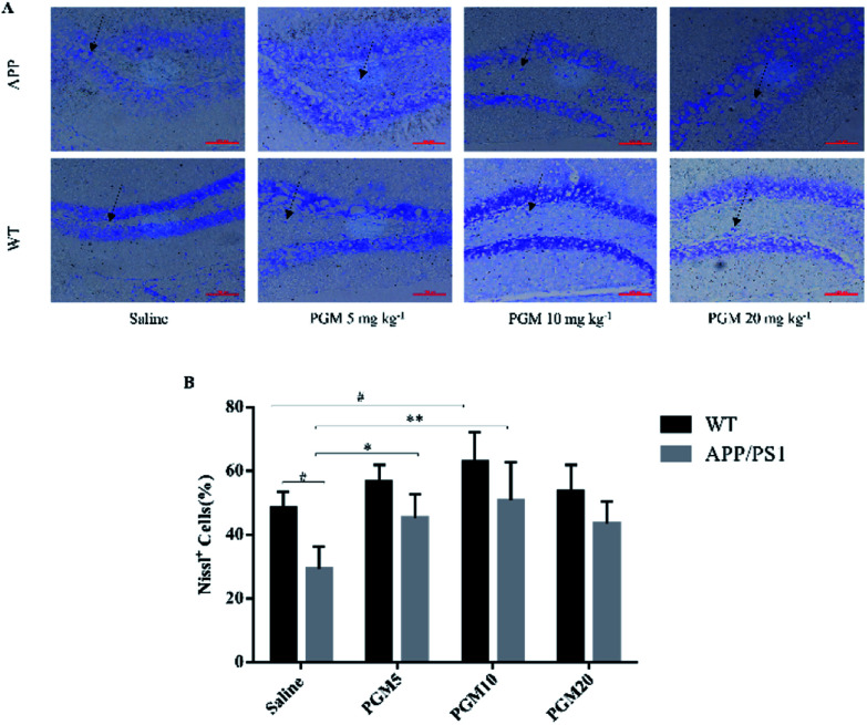
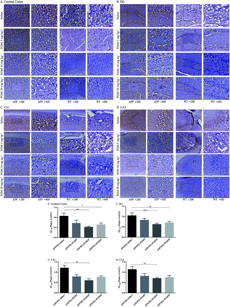
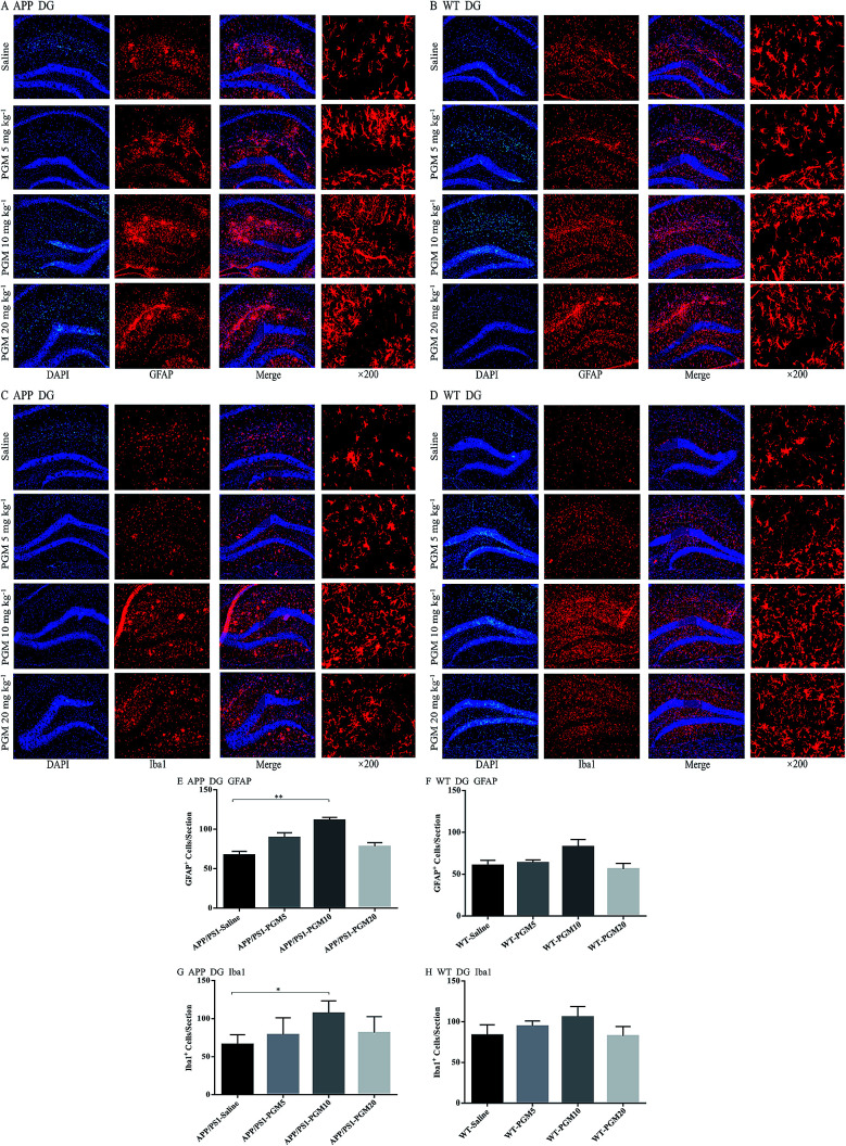
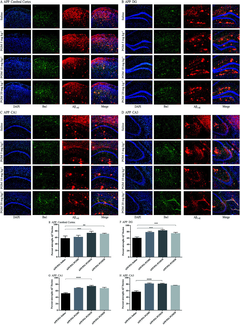
Alzheimer's disease (AD) is characterized by the deposition of amyloid-β (Aβ) plaques, neuronal loss and neurofibrillary tangles. In addition, neuroinflammatory processes are thought to contribute to AD pathophysiology. Maitake (Grifola frondosa), an edible/medicinal mushroom, exhibits high nutritional value and contains a great amount of health-beneficial, bioactive compounds. It has been reported that proteo-β-glucan, a polysaccharide derived from Maitake (PGM), possesses strong immunomodulatory activities. However, whether PGM is responsible for the immunomodulatory and neuroprotection effects on APPswe/PS1ΔE9 (APP/PS1) transgenic mice, a widely used animal model of AD, remains unclear. In the present study, the results demonstrated that PGM could improve learning and memory impairment, attenuate neuron loss and histopathological abnormalities in APP/PS1 mice. In addition, PGM treatment could activate microglia and astrocytes and promote microglial recruitment to the Aβ plaques. Also, PGM could enhance Aβ phagocytosis, and thereby alleviate Aβ burden and the pathological changes in the cortex and hippocampus in APP/PS1 mice. Moreover, PGM showed no significant effect on mice body weight. In conclusion, these findings indicated that administration of PGM could improve memory impairment via immunomodulatory action, and dietary supplementation with PGM may provide potential benefits on brain aging related memory dysfunction.


Conflict of interest statement

There are no conflicts to declare.

Figures

Fig. 1
Fig. 1. Schematic representation of the experimental design.
Fig. 2
Fig. 2. PGM ameliorated learning and memory deficits in APP/PS1 mice in Morris water maze test. n = 12 mice per group. (A) The changes of swimming path. (B and C) The latency to find a hidden platform in APP/PS1 (B) and WT mice (C). (D and E) The number of platform crossing during the spatial probe test in APP/PS1 (D) and WT mice (E). #P < 0.05 vs. WT-saline, *P < 0.05 vs. APP/PS1-saline.
Fig. 3
Fig. 3. H&E staining of neuronal morphology in hippocampal regions (include DG, CA1 and CA3). (A–C) Representative images of APP/PS1 mice groups. (D and E) Representative images of WT mice groups. Scale bar = 100 μm. n = 5–8 mice per group.
Fig. 4
Fig. 4. Nissl staining of neuronal morphology in hippocampal DG region. (A) Representative light micrographs of Nissl-stained DG hippocampal cells. Scale bar = 100 μm. (B) Quantitative analysis of Nissl cells in the DG regions of the hippocampus. n = 5–8 mice per group. #P < 0.05 vs. WT-saline, *P < 0.05 vs. APP/PS1-saline, **P < 0.01 vs. APP/PS1-saline.
Fig. 5
Fig. 5. PGM alleviated brain Aβ1-42 burden in APP/PS1 mice. (A–D) Respectively representative images of Aβ1-42 deposits in the cerebral cortex and the DG, CA1, CA3 regions of the hippocampus. Scale bar = 100 μm. (E–H) Respectively quantification of Aβ1-42 area in the cerebral cortex and the DG, CA1, CA3 regions of the hippocampus. n = 5–8 mice per group, three sections per animal. *P < 0.05 vs. APP/PS1-saline, **P < 0.01 vs. APP/PS1-saline, ***P < 0.001 vs. APP/PS1-saline.
Fig. 6
Fig. 6. PGM aggrandized glial cell activation in the hippocampal regions of APP/PS1 mice. (A) Fluorescent GFAP (red)/DAPI (blue) colocalization in the DG regions of the hippocampus in APP/PS1 mice. Scale bar = 100 μm. (B) Fluorescent GFAP (red)/DAPI (blue) colocalization in the DG regions of the hippocampus in WT mice. Scale bar = 100 μm. (C) Fluorescent Iba1 (red)/DAPI (blue) colocalization in the DG regions of the hippocampus in APP/PS1 mice. Scale bar = 100 μm. (D) Fluorescent Iba1 (red)/DAPI (blue) colocalization in the DG regions of the hippocampus in WT mice. Scale bar = 100 μm. (E) Quantification of GFAP-positive cells in each APP/PS1 mice group. n = 5–8 per group, three sections per animal. (F) Quantification of GFAP-positive cells in each WT mice group. n = 5–8 per group, three sections per animal. (G) Quantification of Iba1-positive cells in each APP/PS1 mice group. n = 5–8 per group, three sections per animal. (H) Quantification of Iba1-positive cells in each WT mice group, n = 5–8 per group, three sections per animal. *P < 0.05 vs. APP/PS1-saline, **P < 0.01 vs. APP/PS1-saline.
Fig. 7
Fig. 7. PGM aggrandized glial cell activation and increased amyloid endocytosis by microglia in the hippocampal regions of APP/PS1 mice. (A–D) Fluorescent Aβ1-42 (red)/Iba1 (green)/DAPI (blue) colocalization in the cortex (A) and the DG (B), CA1 (C), CA3 (D) regions of the hippocampus in APP/PS1 mice. Scale bar = 100 μm. (E–H) Percentage of Iba1-positive microglia cell bodies that are also Aβ-positive in the cortex (E) and the DG (F), CA1 (G) and CA3 (H) regions of the hippocampus. n = 5–8 mice per group, three sections per animal. **P < 0.01 vs. APP/PS1-saline, ***P < 0.001 vs. APP/PS1-saline, ****P < 0.0001 vs. APP/PS1-saline.

References

    1. Pan R. Y. Ma J. Kong X. X. Wang X. F. Li S. S. Qi X. L. Yan Y. H. Cheng J. Liu Q. Jin W. Tan C. H. Yuan Z. Sodium rutin ameliorates Alzheimer's disease-like pathology by enhancing microglial amyloid-beta clearance. Sci. Adv. 2019;5:eaau6328. doi: 10.1126/sciadv.aau6328. - DOI - PMC - PubMed
    1. Zhu D. Yang N. Liu Y. Y. Zheng J. Ji C. Zuo P. P. M2 Macrophage Transplantation Ameliorates Cognitive Dysfunction in Amyloid-beta-Treated Rats Through Regulation of Microglial Polarization. J. Alzheimer's Dis. 2016;52:483–495. - PubMed
    1. Heneka M. T. Golenbock D. T. Latz E. Innate immunity in Alzheimer's disease. Nat. Immunol. 2015;16:229–236. doi: 10.1038/ni.3102. - DOI - PubMed
    1. Hardy J. Selkoe D. J. The amyloid hypothesis of Alzheimer's disease: progress and problems on the road to the rapeutics. Science. 2002;297:353–356. doi: 10.1126/science.1072994. - DOI - PubMed
    1. Mucke L. Selkoe D. J. Neurotoxicity of amyloid beta-protein: synaptic and network dysfunction. Cold Spring Harbor Perspect. Med. 2012;2:a006338. - PMC - PubMed