. 2018 Jan 8;8(4):1730-1736.
doi: 10.1039/c7ra12449j. eCollection 2018 Jan 5.

A three-dimensional hydroxyapatite/polyacrylonitrile composite scaffold designed for bone tissue engineering

Affiliations

A three-dimensional hydroxyapatite/polyacrylonitrile composite scaffold designed for bone tissue engineering

Shuyi Wu et al. RSC Adv. .

Abstract

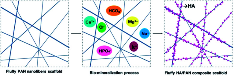
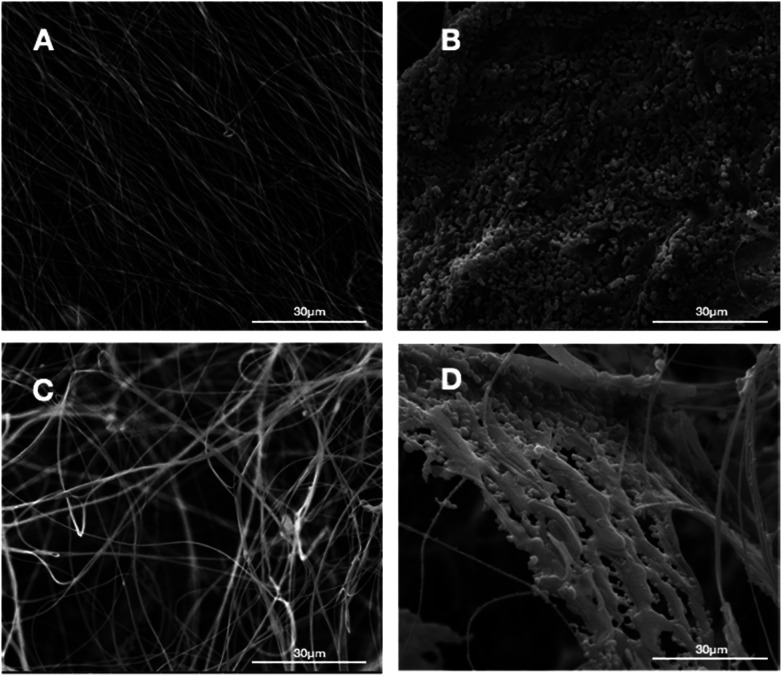
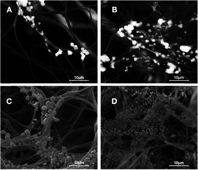
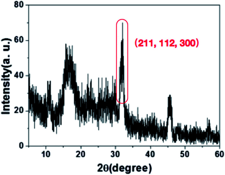
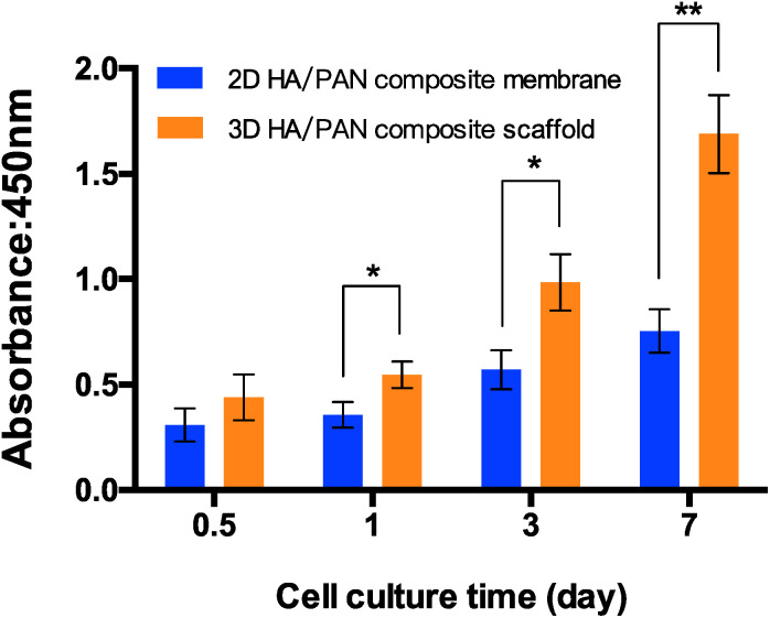
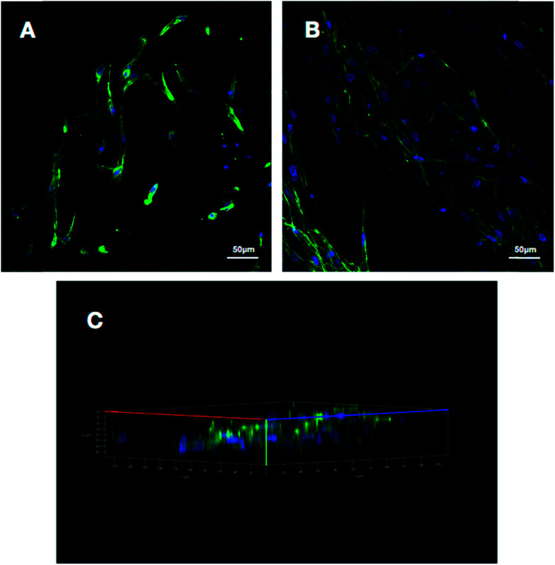
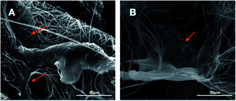
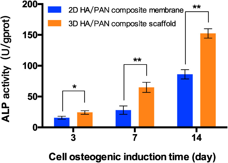
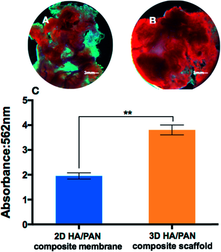
In recent years, various composite scaffolds based on hydroxyapatite have been developed for bone tissue engineering. However, the poor cell survival micro-environment is still the major problem limiting their practical applications in bone repairing and regeneration. In this study, we fabricated a class of fluffy and porous three-dimensional composite fibrous scaffolds consisting of hydroxyapatite and polyacrylonitrile by employing an improved electrospinning technique combined with a bio-mineralization process. The fluffy structure of the hydroxyapatite/polyacrylonitrile composite scaffold ensured the cells would enter the interior of the scaffold and achieve a three-dimensional cell culture. Bone marrow mesenchymal stem cells were seeded into the scaffolds and cultured for 21 days in vitro to evaluate the response of cellular morphology and biochemical activities. The results indicated that the bone marrow mesenchymal stem cells showed higher degrees of growth, osteogenic differentiation and mineralization than those cultured on the two-dimensional hydroxyapatite/polyacrylonitrile composite membranes. The obtained results strongly supported the fact that the novel three-dimensional fluffy hydroxyapatite/polyacrylonitrile composite scaffold had potential application in the field of bone tissue engineering.


Conflict of interest statement

There are no conflicts to declare.

Figures

Fig. 1
Fig. 1. Fabrication process of 3D HA/PAN composite scaffold.
Fig. 2
Fig. 2. SEM images of PAN membrane and fluffy PAN scaffold before and after mineralization of HA: (A) 2D PAN membrane, (B) 2D HA/PAN composite membrane, (C) 3D PAN scaffold, and (D) 3D HA/PAN composite scaffold.
Fig. 3
Fig. 3. SEM images of the 3D HA/PAN composite scaffold after incubation in SBF at different time points: (A) day 3, (B) day 7, (C) day 14, (D) day 28.
Fig. 4
Fig. 4. XRD of 3D HA/PAN composite scaffold.
Fig. 5
Fig. 5. The cytotoxicity and proliferation of BMSCs cultured on 2D HA/PAN composite membrane and 3D HA/PAN composite scaffold at various time points by CCK-8 assay; data are mean ± SD, n = 4, *P < 0.05, **P < 0.01.
Fig. 6
Fig. 6. Fluorescence images of BMSCs cultured on different scaffolds at day 3: (A) 2D HA/PAN composite membrane, (B) 3D HA/PAN composite scaffold and (C) three-dimensional reconstruction of 3D HA/PAN composite scaffold cells.
Fig. 7
Fig. 7. SEM images of BMSCs cultured on different scaffolds at day 3: (A) 2D HA/PAN composite membrane and (B) 3D HA/PAN composite scaffold; yellow arrow: BMSCs.
Fig. 8
Fig. 8. ALP activity of BMSCs cultured on 2D HA/PAN composite membrane and 3D HA/PAN composite scaffold; data are mean ± SD, n = 4, *P < 0.05, **P < 0.01.
Fig. 9
Fig. 9. Alizarin red S staining of BMSCs cultured on 2D HA/PAN composite membrane (A) and 3D HA/PAN composite scaffold (B); semi-quantitatively analysis of calcium content (C); data are mean ± SD, n = 4, **P < 0.01.

References

    1. Fratzl P. Weinkamer R. Prog. Mater. Sci. 2007;52:1263–1334. doi: 10.1016/j.pmatsci.2007.06.001. - DOI
    1. D'Mello S. Elangovan S. Hong L. Ross R. D. Sumner D. R. Salem A. K. J. Biomed. Mater. Res., Part B. 2015;103:1044–1049. doi: 10.1002/jbm.b.33290. - DOI - PMC - PubMed
    1. Fountain S. Windolf M. Henkel J. Tavakoli A. Schuetz M. A. Hutmacher D. W. Epari D. R. Tissue Eng., Part B. 2016;22:47–57. - PubMed
    1. Jeon O. H. Elisseeff J. Drug Delivery Transl. Res. 2016;6:105–120. doi: 10.1007/s13346-015-0266-7. - DOI - PubMed
    1. Kokubo T. Kim H. M. Kawashita M. Biomaterials. 2003;24:2161–2175. doi: 10.1016/S0142-9612(03)00044-9. - DOI - PubMed