Review
. 2018 Jan 9;8(4):2015-2033.
doi: 10.1039/c7ra11278e. eCollection 2018 Jan 5.

Biological properties of calcium phosphate biomaterials for bone repair: a review

Affiliations
Review

Biological properties of calcium phosphate biomaterials for bone repair: a review

Jingyi Lu et al. RSC Adv. .

Abstract

Bone defects are a common disease threatening the health of many people. Calcium phosphate (CaP) is an ideal bone substitutive material that is widely used for bone repair due to its excellent biological properties including osteoinductivity, osteoconductivity and biodegradability. For this reason, investigation of these properties and the effects of various influencing factors is vital for modulating calcium phosphate during the design process to maximally satisfy clinical requirements. In this study, the latest studies on the biological properties of CaP biomaterials, including hydroxyapatite (HA), tricalcium phosphate (TCP), and biphasic calcium phosphate (BCP), have been summarized. Moreover, recent advances on how these properties are altered by different factors are reviewed. Considering the limited mechanical strength of CaP materials, this study also reviews CaP composites with different materials as improvement measures. Finally, perspectives regarding future developments of CaP materials are also provided.


Conflict of interest statement

There are no conflicts to declare.

Figures

Fig. 1
Fig. 1. TGF-β signaling and negative regulation in bone formation. Reprinted with permission from ref. 41 (Copyright © 2012, IVYSPRING INTERNATIONAL PUBLISHER).
Fig. 2
Fig. 2. Extracellular regulators of the Wnt signaling pathway: (A) Wnt ligands use diverse co-receptors to activate and modulate different downstream signals in the Wnt signaling pathway. (B) After the binding of Wnt ligands to the frizzled receptor and LRPs co-receptors, Wnt signaling is activated and causes the transcription of gene targets. (C) Models of Wnt signaling inhibition. Reprinted with permission from ref. 46 (Copyright © 2017, Elsevier).
Fig. 3
Fig. 3. Scanning electron microscopy technique: (a) stem cells adhered in the pores of the scaffold and (b) extracellular matrix as the deposition of granular products secreted by differentiated osteoblasts in the pores of the hydroxyapatite/tricalcium phosphate scaffold. Reprinted with permission from ref. 59 (Copyright © 2016, Wolters Kluwer India Pvt. Ltd).
Fig. 4
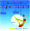
Fig. 4. Schematic of the vascularization strategy within the channels of the CaP scaffold: (a) different channel diameters induced different expression behaviors for growth factors and then induced the different vessel formation; (b) the gradually-increased HIF1α expression in the channels induced the in-growth of blood vessels into its host. Reprinted with permission from ref. 62 (Copyright © 2016, Elsevier).
Fig. 5
Fig. 5. Osteoclasts function on the surface of CaP. Reprinted with permission from ref. 80 (Copyright © 2017, Elsevier).
Fig. 6
Fig. 6. A scanning electron micrograph showing bone formation within (a) the SiCaP-20 scaffold; (b) the SiCaP-30 scaffold. Reprinted with permission from ref. 97 (Copyright © 2012, John Wiley and Sons).
Fig. 7
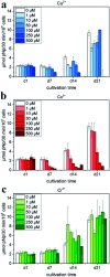
Fig. 7. ALP activity of hMSC cultivated in the presence of Cu2+, Co2+ and Cr3+ ions added to cell culture medium (with OS) in various concentrations: (a) Cu2+: 0–500 μM (μmol L−1); (b) Co2+: 0–500 μM; and (c) Cr3+: 0–500 μM. Reprinted with permission from ref. 111 (Copyright © 2017, Elsevier).
Fig. 8
Fig. 8. Formation mechanism of apatite deposited on the substrate with PDA coating. Reprinted with permission from ref. 128 (Copyright © 2010, John Wiley and Sons).
Fig. 9
Fig. 9. Schematic showing the apparatus for studying the degradation of scaffolds under conditions of fluid flow. Reprinted with permission from ref. 141 (Copyright © 2007, Trans Tech Publications).
Fig. 10
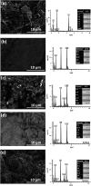
Fig. 10. SEM and the corresponding EDX measurements on the Ti–6Al–4V substrate (a) on a CaP coating (b), on a Ag_CaP coating (c), on a Zn_CaP coating (d) as well as on a AgZn_CaP coating (e). Reprinted with permission from ref. 149 (Copyright © 2016, Elsevier).
Fig. 11
Fig. 11. (Top) After 1 month of healing, (A) there was no bone formation around the defect in the negative control. (B and C) For the PMMA–brushite cement, osseointegration was observed with initial bone remodeling indicated by the formation of a cutting cone (area highlighted in the yellow square in (B), and magnified in (C)). (Bottom) The histology slides are shown for the cement test groups after 2 months of healing. Bone formation was only observed around the composite (E) PMMA–HA and (F) PMMA–brushite cements with no bone formation shown around (D) PMMA–K. Reprinted with permission from ref. 162 (Copyright © 2017, American Chemical Society).
None
Jingyi Lu
None
Huijun Yu
None
Chuanzhong Chen

References

    1. Denry I. Kuhn L. T. Dent. Mater. 2016;32:43–53. doi: 10.1016/j.dental.2015.09.008. - DOI - PMC - PubMed
    1. Shao R. Quan R. Zhang L. Wei X. Yang D. Xie S. J. Ceram. Soc. Jpn. 2015;123:17–20. doi: 10.2109/jcersj2.123.17. - DOI
    1. Lee E. J. Kasper F. K. Mikos A. G. Ann. Biomed. Eng. 2014;42:323–337. doi: 10.1007/s10439-013-0859-6. - DOI - PMC - PubMed
    1. Liu C. Tang R. Chin. J. Inorg. Chem. 2014;30:1–9.
    1. Tang Z. Tan Y. Ni Y. Wang J. Zhu X. Fan Y. Chen X. Yang X. Zhang X. Mater. Sci. Eng., C. 2017;70:1000–1010. doi: 10.1016/j.msec.2016.06.097. - DOI - PubMed