Review
. 2018 Jan 19;8(7):3736-3749.
doi: 10.1039/c7ra11593h. eCollection 2018 Jan 16.

Chitosan composite scaffolds for articular cartilage defect repair: a review

Affiliations
Review

Chitosan composite scaffolds for articular cartilage defect repair: a review

Huijun Li et al. RSC Adv. .

Abstract

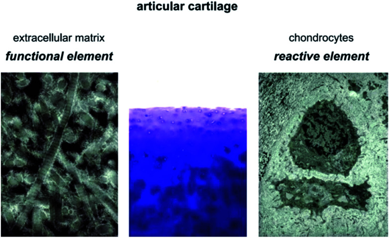
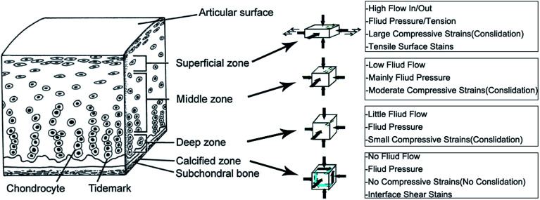
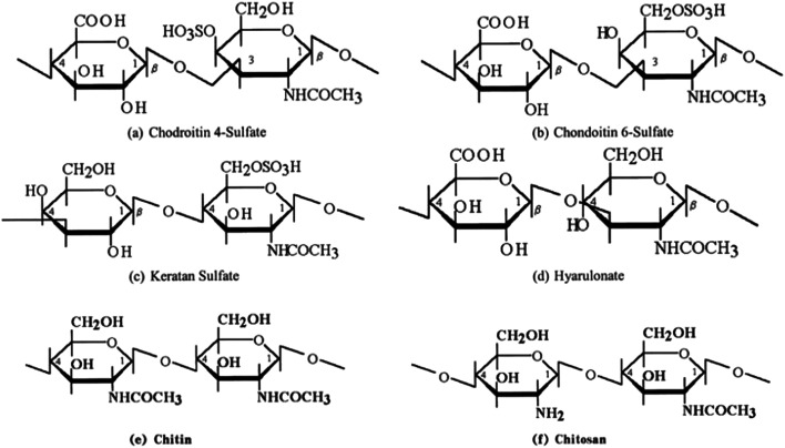
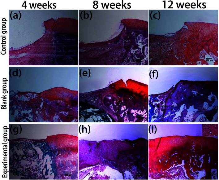
Articular cartilage (AC) defects lack the ability to self-repair due to their avascular nature and the declined mitotic ability of mature chondrocytes. To date, cartilage tissue engineering using implanted scaffolds containing cells or growth factors is the most promising defect repair method. Scaffolds for cartilage tissue engineering have been comprehensively researched. As a promising scaffold biomaterial for AC defect repair, the properties of chitosan are summarized in this review. Strategies to composite chitosan with other materials, such as polymers (including collagen, gelatin, alginate, silk fibroin, poly-caprolactone, and poly-lactic acid) and bioceramics (including calcium phosphate, calcium polyphosphate, and hydroxyapatite) are presented. Methods to manufacture three-dimensional porous structures to support cell attachment and nutriment exchange have also been included.


Conflict of interest statement

There are no conflicts to declare.

Figures

Fig. 1
Fig. 1. Constitution of articular cartilage.
Fig. 2
Fig. 2. Structure of adult articular cartilage and the in vivo mechanical environment.
Fig. 3
Fig. 3. Molecular structures of some polysaccharide repeating units and chitosan.
Fig. 4
Fig. 4. Treatment of Enterobacter aerogenes and Staphylococcus aureus with different types of chitosan solution.
Fig. 5
Fig. 5. SEM micrographs of chitosan microcarriers adhered to different growth progressions of human skin fibroblast on the bead surface. (a) Fibroblast after 24 h of incubation. (b) Fibroblast after 120 h of incubation.
Fig. 6
Fig. 6. Biocompatibility of MNPs and CH-MNPs. Cell viability data for MNPs and CH-MNPs for 24 and 48 h on L929, HeLa, and MCF7 cells. Values are expressed as mean ± SD (n = 3), LSMO (La0.7Sr0.3MnO3).
Fig. 7
Fig. 7. SEM images of chondrocyte cells grown on (a) chitosan–alginate and (b) chitosan scaffolds after 14 days of cell culture.
Fig. 8
Fig. 8. (a) Degradation behavior and (b) change in pH of solutions of SF/CS blended scaffolds in 0.05 M PBS containing 1.6 μg mL−1 of lysozyme or pure PBS (pH 7.4) at 37 °C.
Fig. 9
Fig. 9. Wright's stain images of cells grown on scaffolds (original magnification, 200×). (a) HA/CS composite scaffolds, (b) chitosan scaffolds. According to the reference cited, (a) and (b) have the same original size of 5.87 cm × 7.32 cm.
Fig. 10
Fig. 10. Mechanical properties of repair tissues in different groups: (a) elastic modulus of repair tissue determined by nanoindentation; (b) hardness of repair tissue determined by nanoindentation.
Fig. 11
Fig. 11. Morphology of porous chitosan scaffold with pores sizes of (a) ≤10 μm, (b) 10–50 μm, and (c) 70–120 μm in diameter. Scale bar = 100 μm.
Fig. 12
Fig. 12. Gross morphology of repaired cartilage in (a–c) the control group, (d–f) the blank group, and (g–i) the experimental group at 4, 8, and 12 weeks.
Fig. 13
Fig. 13. Safranin-O staining of repaired cartilage in (a–c) the control group, (d–f) the blank group, and (g–i) the experimental group at 4, 8, and 12 weeks. Scale bar = 200 μm. Arrows show chitosan scaffold.
None
Huijun Li
None
Cheng Hu
None
Huijun Yu
None
Chuanzhong Chen

Similar articles

Cited by

References

    1. Duarte Campos D. F. Drescher W. Rath B. Tingart M. Fischer H. Cartilage. 2012;3:205–221. doi: 10.1177/1947603512444722. - DOI - PMC - PubMed
    1. Ebihara G. Sato M. Yamato M. Mitani G. Kutsuna T. Nagai T. Ito S. Ukai T. Kobayashi M. Kokubo M. Okano T. Mochida J. Biometerials. 2012;33:3846–3851. doi: 10.1016/j.biomaterials.2012.01.056. - DOI - PubMed
    1. Makris E. A. Gomoll A. H. Malizos K. N. Hu J. C. Athanasiou K. A. Nat. Rev. Rheumatol. 2015;11:21–34. doi: 10.1038/nrrheum.2014.157. - DOI - PMC - PubMed
    1. Griffith L. G. Naughton G. Science. 2002;295:1009–1016. doi: 10.1126/science.1069210. - DOI - PubMed
    1. Kock L. van Donkelaar C. C. Ito K. Cell Tissue Res. 2012;347:613–627. doi: 10.1007/s00441-011-1243-1. - DOI - PMC - PubMed