

doi: 10.1039/c7ra13499a.
eCollection 2018 Jan 16.
Eugenol triggers CD11b+Gr1+ myeloid-derived suppressor cell apoptosis via endogenous apoptosis pathway
Affiliations
- PMID: 35542913
- PMCID: PMC9077712
- DOI: 10.1039/c7ra13499a
Item in Clipboard
Eugenol triggers CD11b+Gr1+ myeloid-derived suppressor cell apoptosis via endogenous apoptosis pathway
RSC Adv.
.
Abstract
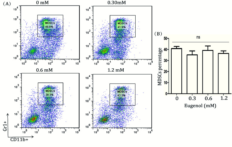
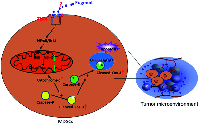
To study the effect and underlying molecular mechanism of eugenol on CD11b+Gr1+ myeloid-derived suppressor cells (MDSCs). The effect of eugenol on the inhibition of immortalized MDSC cell line MSC-2 and murine peritoneal macrophages was detected by MTT. Flow cytometry was used to detect the pro-apoptosis effect of eugenol on MDSCs. The expression levels of apoptosis-related proteins were detected by western blot. Eugenol has a selective inhibitory effect on MDSCs in a dose-dependent manner, which activates an endogenous apoptosis pathway, leading to apoptosis. Eugenol promotes the apoptosis of MDSCs via the intrinsic pathway.
This journal is © The Royal Society of Chemistry.
Conflict of interest statement
There are no conflicts to declare.
Figures


Similar articles
-
Microsomal prostaglandin E synthase-1 promotes lung metastasis via SDF-1/CXCR4-mediated recruitment of CD11b+Gr1+MDSCs from bone marrow.Biomed Pharmacother. 2020 Jan;121:109581. doi: 10.1016/j.biopha.2019.109581. Epub 2019 Nov 10. Biomed Pharmacother. 2020. PMID: 31715374
-
miR-223 suppresses differentiation of tumor-induced CD11b⁺ Gr1⁺ myeloid-derived suppressor cells from bone marrow cells.Int J Cancer. 2011 Dec 1;129(11):2662-73. doi: 10.1002/ijc.25921. Epub 2011 Mar 25. Int J Cancer. 2011. PMID: 21213211
-
Decidua-derived granulocyte macrophage colony-stimulating factor induces polymorphonuclear myeloid-derived suppressor cells from circulating CD15+ neutrophils.Hum Reprod. 2020 Dec 1;35(12):2677-2691. doi: 10.1093/humrep/deaa217. Hum Reprod. 2020. PMID: 33067638
-
Myeloid-derived suppressor cells in breast cancer.Breast Cancer Res Treat. 2013 Jul;140(1):13-21. doi: 10.1007/s10549-013-2618-7. Epub 2013 Jul 5. Breast Cancer Res Treat. 2013. PMID: 23828498 Free PMC article. Review.
-
The roles of myeloid-derived suppressor cells in transplantation.Expert Rev Clin Immunol. 2014 Oct;10(10):1385-94. doi: 10.1586/1744666X.2014.948424. Epub 2014 Aug 13. Expert Rev Clin Immunol. 2014. PMID: 25119260 Review.
Cited by
-
In Vitro Anti-Leishmanial Activity and Molecular Docking of Eugenol as a Potential Agent Against Leishmania Major.Acta Parasitol. 2025 Jul 7;70(4):147. doi: 10.1007/s11686-025-01089-w. Acta Parasitol. 2025. PMID: 40622619
-
Ligustilide Inhibits Tumor Angiogenesis by Downregulating VEGFA Secretion from Cancer-Associated Fibroblasts in Prostate Cancer via TLR4.Cancers (Basel). 2022 May 13;14(10):2406. doi: 10.3390/cancers14102406. Cancers (Basel). 2022. PMID: 35626012 Free PMC article.
References
LinkOut - more resources
Full Text Sources
Research Materials