. 2022 Jun;25(6):214.
doi: 10.3892/mmr.2022.12730. Epub 2022 May 11.

Curcumin improves the function of umbilical vein endothelial cells by inhibiting H2O2‑induced pyroptosis

Affiliations

Curcumin improves the function of umbilical vein endothelial cells by inhibiting H2O2‑induced pyroptosis

Yulin Yuan et al. Mol Med Rep. 2022 Jun.

Abstract

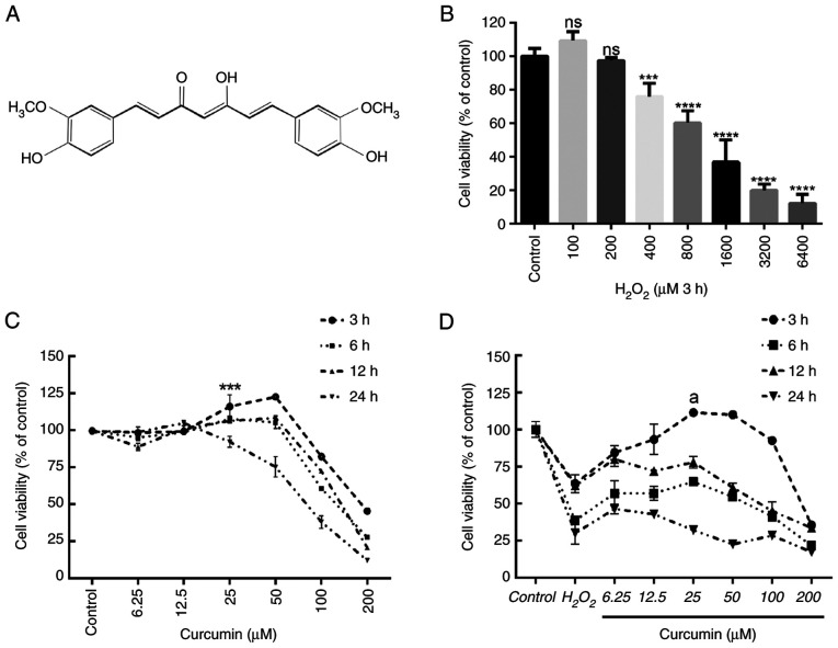
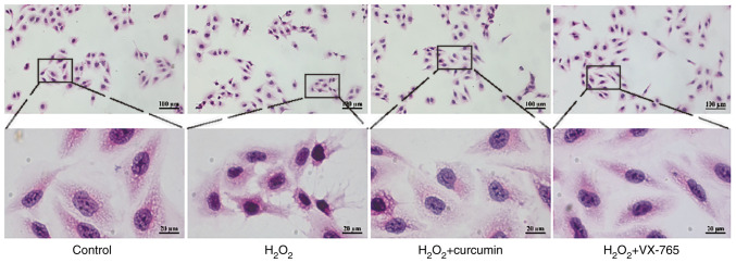
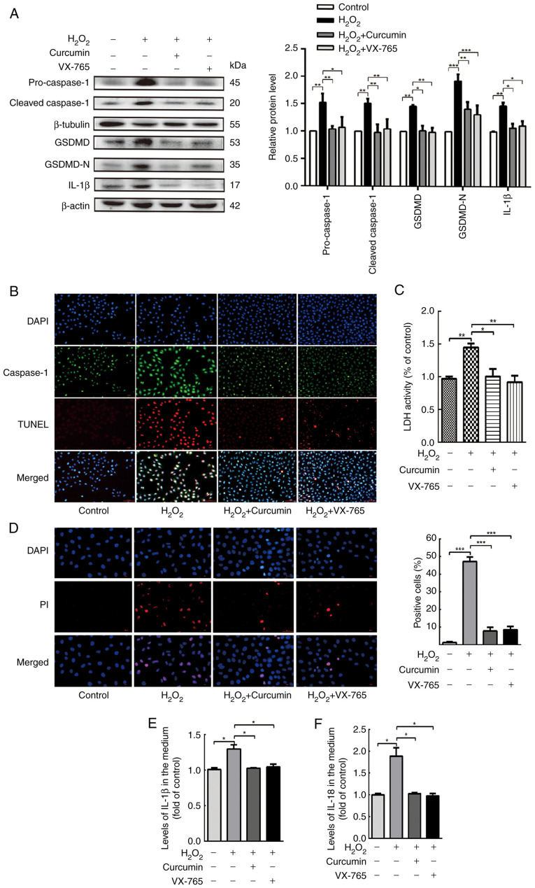
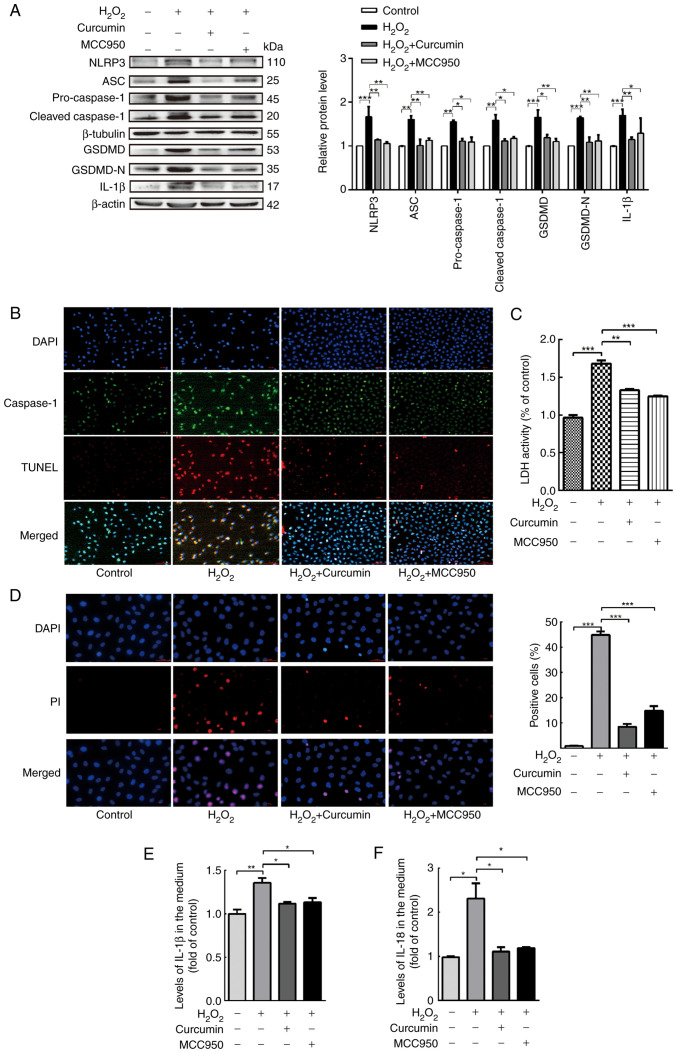
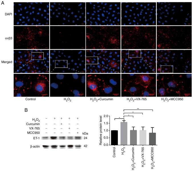
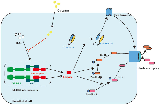
Endothelial cell (EC) dysfunction is one of the initiating factors of atherosclerosis. EC dysfunction is primarily caused by oxidative damage and inflammation. As a classic non‑specific antioxidant and anti‑inflammatory drug, curcumin has been widely used in studies of lipid metabolism disorders. However, whether curcumin is able to alleviate H2O2‑induced EC damage and its related mechanisms has remained to be elucidated. The present study confirmed the protective effects of curcumin on human umbilical vein endothelial cells (HUVECs). A HUVEC injury model was established using H2O2 and the optimal concentrations and time of curcumin to achieve therapeutic effects were explored. Curcumin was observed to inhibit H2O2‑induced pyroptosis by inhibiting the activation of NOD‑, LRR‑ and pyrin domain‑containing protein 3. In addition, curcumin improved HUVEC function by restoring αvβ3 and reducing endothelin‑1 expression. In conclusion, the results of the present study revealed the mechanism through which curcumin inhibits pyroptosis and indicated that curcumin may have a potential utility in treating diseases of EC dysfunction.

Keywords: atherosclerosis; curcumin; endothelial cell; hydrogen peroxide; pyroptosis.


Conflict of interest statement

The authors declare that they have no competing interests.

Figures

Figure 1.
Figure 1.
Curcumin suppresses H2O2-induced viability impairment of HUVECs. The effects of increasing concentrations of H2O2 and curcumin on HUVEC viability were determined. (A) Curcumin contains a variety of functional groups, including the β-diketo group, carbon-carbon double bonds and phenyl rings containing varying amounts of hydroxyl and methoxy substituents. Viability of HUVECs treated with increasing doses of (B) H2O2 (0–6,400 µM) for 3 h and (C) curcumin (0–200 µM) for 3, 6, 12 or 24 h, and (D) different concentrations of curcumin (0–200 µM) for 3, 6, 12 or 24 h after 3 h of pretreatment with H2O2 (800 µM). Cell viability was detected using an MTT assay. Values are expressed as the mean ± standard deviation of three independent experiments. ***P<0.001, ****P<0.0001 vs. the control group; aP<0.001 vs. the H2O2 group; ns, no significance. HUVECs, human umbilical vein endothelial cells.
Figure 2.
Figure 2.
Curcumin repairs H2O2-induced HUVEC damage, as indicated by H&E staining. HUVECs were treated with H2O2 for 3 h and then either treated with 25 µM curcumin for 3 h or left untreated (control), whereas in the VX-765 group, HUVECs were pretreated with caspase-1 inhibitor VX-765 (10 µM) for 1 h and then incubated with H2O2 (800 µM) for 3 h (magnification, ×200; scale bar, 100 µm). The areas indicated by the boxes are enlarged regions for each experimental condition (magnification, ×1,000; scale bar, 20 µm). HUVECs, human umbilical vein endothelial cells.
Figure 3.
Figure 3.
Curcumin suppresses H2O2-induced pyroptosis of HUVECs. HUVECs were treated with H2O2 for 3 h and then either treated with 25 µM curcumin for 3 h or left untreated (control), whereas for the VX-765 group, HUVECs were pretreated with caspase-1 inhibitor VX-765 (10 µM) for 1 h and then incubated with H2O2 (800 µM) for 3 h. (A) Western blot analysis revealed that the protein levels of pro-caspase-1, caspase-1, pro-GSDMD, GSDMD and IL-1β were increased in HUVECs following treatment with H2O2 for 3 h. VX-765 and curcumin inhibited the protein expression of pro-caspase-1, caspase-1, pro-GSDMD, GSDMD and IL-1β. β-actin or β-tubulin was used as an internal control. (B) Compared with the H2O2 group, caspase-1 (green) and TUNEL (red) double-positive cells were decreased in the presence of curcumin or VX-765. The nuclei were stained blue with DAPI (magnification, ×400; scale bar, 50 µm). (C) The relative release of LDH in H2O2-treated HUVECs was increased, while that in curcumin- and VX-765-treated HUVECs was decreased (n=3). (D) The percentage of PI-positive HUVECs (red) was increased following H2O2 treatment, while it decreased following curcumin and VX-765 treatment (left, representative images; right, quantification of PI-positive cells; magnification, ×400; scale bar, 50 µm). Relative concentration of (E) IL-1β and (F) IL-18 in the culture medium following the treatment of HUVECs with H2O2, curcumin or VX-765, as determined by ELISA. Values are expressed as the mean ± standard deviation from three independent experiments. *P<0.05, **P<0.01 and ***P<0.001 as indicated. HUVECs, human umbilical vein endothelial cells; GSDMD, gasdermin D; LDH, lactate dehydrogenase; PI, propidium iodide.
Figure 4.
Figure 4.
Curcumin alleviates NLRP3 inflammasome activation, which is involved in pyroptosis. HUVECs were treated with H2O2 for 3 h and then either treated with 25 µM curcumin for 3 h or left untreated (control), whereas for the MCC95 group, HUVECs were pretreated with NLRP3 inhibitor (MCC950; 10 µM) for 2 h and then incubated with H2O2 (800 µM) for 3 h. (A) Western blot analysis indicated that the protein levels of NLRP3, ASC, pro-caspase-1, caspase-1, pro-GSDMD, GSDMD and IL-1β were upregulated in HUVECs following treatment with H2O2 for 3 h and that MCC950 and curcumin inhibited the protein expression of NLRP3, ASC, pro-caspase-1, caspase-1, pro-GSDMD, GSDMD and IL-1β. β-Actin or β-tubulin was used as an internal control. (B) As compared with the H2O2 group, caspase-1 (green) and TUNEL (red) double-positive cells were decreased in the presence of curcumin or MCC950. The nuclei were stained blue with DAPI (magnification, ×400; scale bar, 50 µm). (C) The relative release of LDH by H2O2-treated HUVECs was increased, while that of curcumin- and MCC950-treated HUVECs was decreased (n=3). (D) The percentage of PI (red)-positive H2O2-treated HUVECs was increased, while that among curcumin- and MCC950-treated cells was decreased (left, representative images; right, quantification of PI-positive cells) (magnification, ×400; scale bar, 50 µm). Relative concentration of (E) IL-1β and (F) IL-18 in the culture medium following H2O2, curcumin or MCC950 treatment of HUVECs, as determined by ELISA. Values are expressed as the mean ± standard deviation of three independent experiments. *P<0.05, **P<0.01, ***P<0.001. HUVECs, human umbilical vein endothelial cells; NLRP3, NOD-, LRR- and pyrin domain-containing protein 3; GSDMD, gasdermin D; ASC, apoptosis-associated speck-like protein containing a caspase recruitment domain; LDH, lactate dehydrogenase; PI, propidium iodide.
Figure 5.
Figure 5.
Curcumin improves H2O2-induced functional damage in HUVECs. HUVECs were pretreated with caspase-1 inhibitor VX-765 (10 µM) for 1 h or NLRP3 inhibitor (MCC950; 10 µM) for 2 h and then incubated with H2O2 (800 µM) for 3 h. HUVECs were treated with H2O2 for 3 h and then either treated with 25 µM curcumin for 3 h or left untreated (control). (A) αvβ3-positive cells (red) were increased following pretreatment with curcumin and inhibitor. The nuclei were stained blue with DAPI. The regions indicated by boxes are enlarged regions for each experimental condition (scale bars, 10 µm). (B) The protein expression levels of ET-1 were upregulated in HUVECs following treatment with H2O2 for 3 h. VX-765, MCC950 and curcumin inhibited the protein expression of ET-1, as indicated by the western blot analysis. β-actin was used as an internal control. Values are expressed as the mean ± standard deviation of three independent experiments. *P<0.05 and **P<0.01. HUVECs, human umbilical vein endothelial cells; NLRP3, NOD-, LRR- and pyrin domain-containing protein 3; αvβ3, alpha v beta 3; ET-1, endothelin 1.
Figure 6.
Figure 6.
Proposed model of curcumin alleviating H2O2-induced pyroptosis of human umbilical vein endothelial cells. NLRP3, NOD-, LRR- and pyrin domain-containing protein 3; GSDMD, gasdermin D; ASC, apoptosis-associated speck-like protein containing a caspase recruitment domain.

Similar articles

Cited by

References

    1. Lusis AJ. Atherosclerosis. Nature. 2000;407:233–241. doi: 10.1038/35025203. - DOI - PMC - PubMed
    1. Welsh P, Grassia G, Botha S, Sattar N, Maffia P. Targeting inflammation to reduce cardiovascular disease risk: A realistic clinical prospect? Br J Pharmacol. 2017;174:3898–3913. doi: 10.1111/bph.13818. - DOI - PMC - PubMed
    1. Miao EA, Leaf IA, Treuting PM, Mao DP, Dors M, Sarkar A, Warren SE, Wewers MD, Aderem A. Caspase-1-induced pyroptosis is an innate immune effector mechanism against intracellular bacteria. Nat Immunol. 2010;11:1136–1142. doi: 10.1038/ni.1960. - DOI - PMC - PubMed
    1. Miao EA, Rajan JV, Aderem A. Caspase-1-induced pyroptotic cell death. Immunol Rev. 2011;243:206–214. doi: 10.1111/j.1600-065X.2011.01044.x. - DOI - PMC - PubMed
    1. Lin J, Shou X, Mao X, Dong J, Mohabeer N, Kushwaha KK, Wang L, Su Y, Fang H, Li D. Oxidized low density lipoprotein induced caspase-1 mediated pyroptotic cell death in macrophages: implication in lesion instability? PLoS One. 2013;8:e62148. doi: 10.1371/journal.pone.0062148. - DOI - PMC - PubMed

MeSH terms