. 2022 May 1;135(9):jcs259596.
doi: 10.1242/jcs.259596. Epub 2022 May 11.

Mitochondrial antiviral-signalling protein is a client of the BAG6 protein quality control complex

Affiliations

Mitochondrial antiviral-signalling protein is a client of the BAG6 protein quality control complex

Peristera Roboti et al. J Cell Sci. .

Abstract

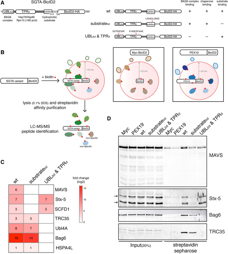
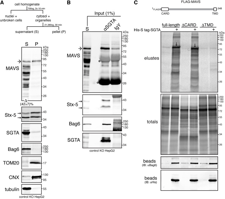
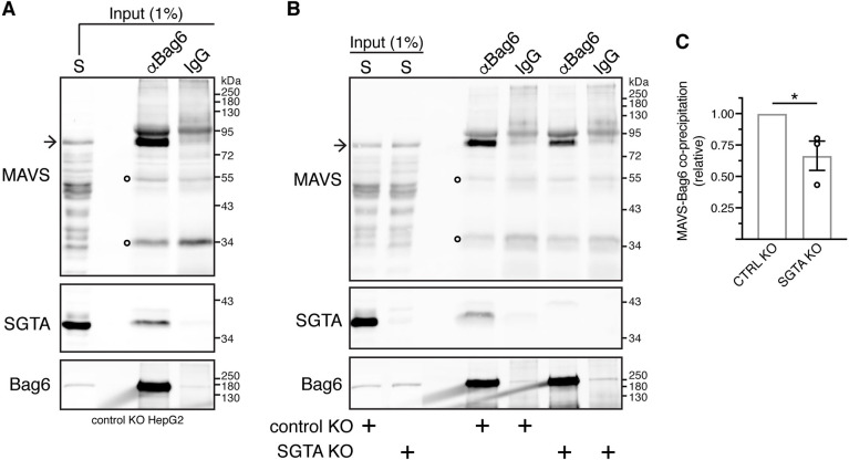
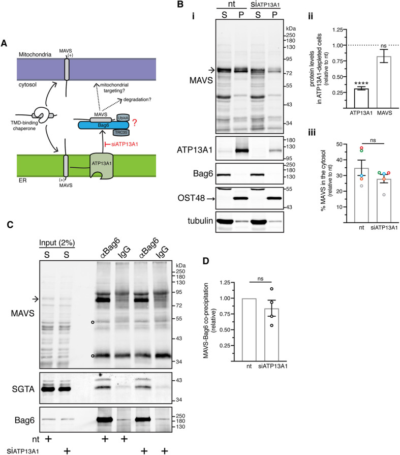
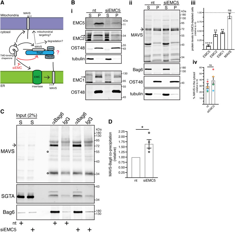
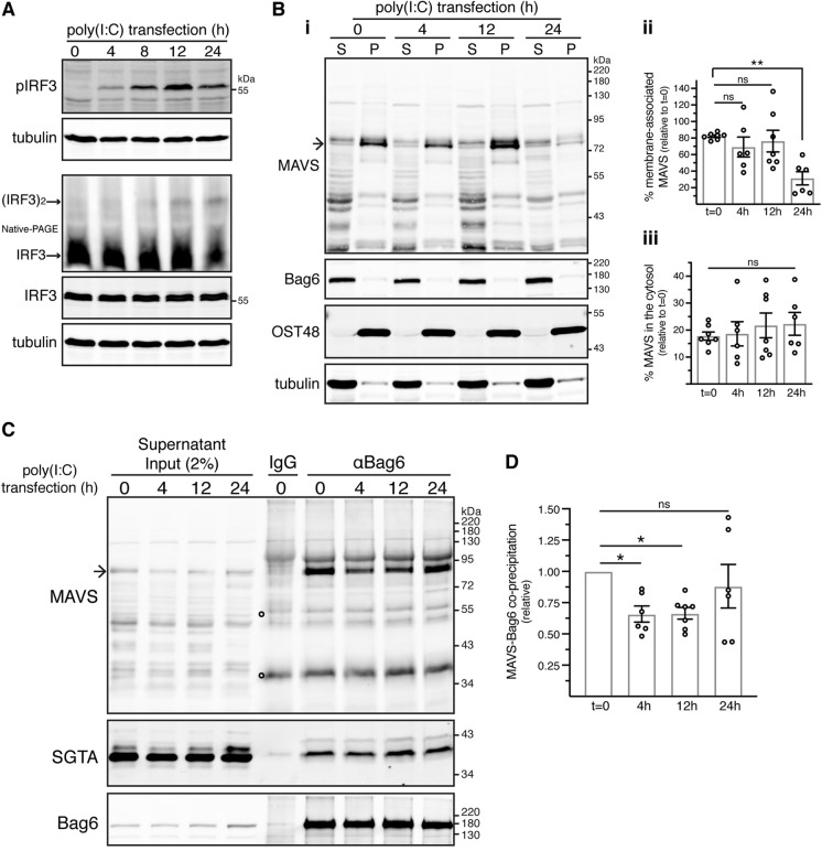
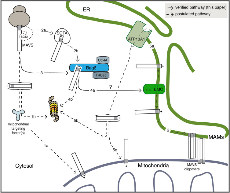
The heterotrimeric BAG6 complex coordinates the direct handover of newly synthesised tail-anchored (TA) membrane proteins from an SGTA-bound preloading complex to the endoplasmic reticulum (ER) delivery component TRC40. In contrast, defective precursors, including aberrant TA proteins, form a stable complex with this cytosolic protein quality control factor, enabling such clients to be either productively re-routed or selectively degraded. We identify the mitochondrial antiviral-signalling protein (MAVS) as an endogenous TA client of both SGTA and the BAG6 complex. Our data suggest that the BAG6 complex binds to a cytosolic pool of MAVS before its misinsertion into the ER membrane, from where it can subsequently be removed via ATP13A1-mediated dislocation. This BAG6-associated fraction of MAVS is dynamic and responds to the activation of an innate immune response, suggesting that BAG6 may modulate the pool of MAVS that is available for coordinating the cellular response to viral infection.

Keywords: BioID2; ER membrane complex; Protein targeting; SGTA; Tail-anchored proteins.


Conflict of interest statement

Competing interests The authors declare no competing or financial interests.

Figures

Fig. 1.
Fig. 1.
MAVS is a high-confidence proximal interactor of SGTA. (A) Left, schematic of human SGTA–BioID2–HA displaying its protein–protein interaction modules. UBLbd, N-terminal domain that binds to the ubiquitin-like (UBL) domains of Ubl4A and Bag6; TPRd, central tetratricopeptide repeat (TPR) domain that interacts with heat-shock proteins; substratebd (sub/tebd), C-terminal domain that contains the hydrophobic substrate-binding site. Right, schematics of the SGTA–BioID2 mutants and respective disrupted interactions. Mutated amino acids are indicated. (B) Scheme for the BioID2-mediated proximity labelling and identification of SGTA substrates and cofactors using SGTA KO cells (see Fig. S1) transiently expressing wild-type (wt) SGTA-BioID2 or mutant variants shown in A. Cells expressing Myc–BioID2 or PEX19–BioID2 serve as two independent controls to exclude non-specific interactors. (C) Heat map representing log2-transformed fold changes in the protein intensities of significant (BFDR<0.05) wild-type (wt)/mutant SGTA-specific preys relative to both the Myc–BioID2 and PEX19–BioID2 controls. Individual rounded values are depicted in the heat map. A non-significant prey is shown as a white box (three biological replicates; see Tables S1–S3 for list of all proteins detected). (D) Validation of selected SGTA-associated candidates from C by immunoblotting. SGTA KO cells expressing the indicated BioID2-tagged baits were treated with biotin for 8 h and lysed with RIPA buffer. The resulting extracts were subjected to affinity purification with streptavidin beads and the bound material eluted using a biotin-containing buffer. The input and eluted material were analysed by immunoblotting for the indicated endogenous proteins. Stx-5 can be observed as two bands (indicated by arrows) corresponding to two isoforms, a 42 kDa-ER and a 35 kDa-Golgi isoform that result from an alternative initiation of translation (Coy-Vergara et al., 2019). Blots representative of three independent biological replicates.
Fig. 2.
Fig. 2.
SGTA interacts with MAVS. (A) A cytosolic pool of endogenous MAVS can be observed at steady-state. Top, schematic of subcellular fractionation protocol used to separate the cell homogenate into crude cytosolic supernatant (S) and membrane-associated pellet (P) fractions. Bottom, detergent-free extracts from control KO cells (see Fig. S1) were fractionated as shown above. Equivalent amounts of each fraction were analysed by immunoblotting for MAVS and various compartmental markers. Bag6, SGTA and tubulin (cytosolic markers), TOM20 (mitochondrial outer membrane marker) and calnexin (CNX, ER membrane marker) serve as fractionation controls. Note that the MAVS-specific antibody, raised against amino acids 1–135 of human MAVS, detected the ∼80 kDa full-length MAVS (marked by an arrow) and multiple shorter variants that most likely represent C-terminally degraded products or processed forms of the full-length protein (see also Seth et al., 2005). Quantification of the levels of full-length MAVS recovered in the cytosolic fraction is indicated below the MAVS blot. Value represents mean±s.e.m. from three independent experiments. (B) MAVS co-immunoprecipitates with SGTA. The supernatant (S) fraction from A was subjected to immunoprecipitations with equal amounts of chicken anti-SGTA antibody (αSGTA) or chicken IgY antibody (control for non-specific binding). Input and immunoprecipitates were analysed by immunoblotting for the indicated endogenous proteins. Bag6 served as positive control for SGTA binding. In A and B, arrows next to the Stx-5 blots indicate the two Stx-5 isoforms. Open circles on MAVS blots indicate signals derived from denatured antibody heavy and light chains. Blots representative of three independent experiments. (C) In vitro translated MAVS interacts with recombinant SGTA via its transmembrane domain (TMD). Top, schematic of FLAG–MAVS displaying its N-terminal caspase activation and recruitment domain (CARD) and C-terminal TMD. Bottom, FLAG-MAVS full-length, ΔCARD or ΔTMD truncated variants were translated in vitro in the absence or presence (+) of 2 µM His-S-tag-SGTA. A 10% sample of the total translation products was subjected to denaturing immunoprecipitations with anti-FLAG antibody (totals), while the rest was incubated with HisPur cobalt resin and bound proteins were eluted using imidazole (eluates). Totals and eluates were resolved by SDS-PAGE and results visualised by phosphorimaging. Downward arrows indicate full-length and ΔCARD FLAG–MAVS selectively bound by His-S-tag-SGTA. His-S-tag-SGTA and its binding partners within rabbit reticulocyte lysate were released from the resin by incubating the beads with SDS sample buffer (beads) and samples were analysed by immunoblotting (IB). The anti-His and anti-Bag6 immunoblots indicate uniform binding of Bag6 binding-competent His-S-tag-SGTA to beads. Results shown in C are representative of two independent experiments.
Fig. 3.
Fig. 3.
Bag6 interacts with MAVS. (A) MAVS co-immunoprecipitates with Bag6. Control KO cells were fractionated as shown in Fig. 2A and the supernatant (S) fraction was subjected to immunoprecipitations with equal amounts of rabbit anti-Bag6 antibody (αBag6) or rabbit IgG antibody (control for non-specific binding). Input and immunoprecipitates were analysed by immunoblotting for the indicated endogenous proteins. SGTA served as positive control for Bag6 binding. (B) SGTA facilitates Bag6–MAVS interaction. Control KO and SGTA KO cells were fractionated as shown in Fig. 2A and the supernatant (S) fractions were subjected to immunoprecipitations with rabbit anti-Bag6 antibody (αBag6) or rabbit control IgG antibody. Inputs and immunoprecipitates were analysed by immunoblotting for the indicated endogenous proteins. SGTA served as positive control for Bag6 binding. Arrow in A and B indicates full-length MAVS. Open circles on MAVS blots indicate signals derived from denatured antibody heavy and light chains. (C) Mean±s.e.m. of MAVS levels that co-immunoprecipitate with Bag6 in control KO and SGTA KO cells for three independent experiments as in B. *P<0.05 (unpaired two-tailed t-test).
Fig. 4.
Fig. 4.
ATP13A1 depletion has no visible effect on Bag6-MAVS interaction. (A) Proposed model. Bag6 recruits MAVS after its ATP13A1-mediated extraction from the ER membrane. Depleting ATP13A1 will decrease Bag6-MAVS interaction, as MAVS cannot be dislocated from the ER membrane. (B) ATP13A1 depletion does not grossly alter the levels of MAVS in the crude cytosolic supernatant fraction. (i) Control KO cells transfected with non-targeting (nt) or ATP13A1-targeting siRNAs (siATP13A1) were fractionated as shown in Fig. 2A. Equivalent amounts of supernatant (S) and pellet (P) fractions were analysed by immunoblotting for the indicated endogenous proteins. (ii) ATP13A1 and MAVS levels in siATP13A1-treated cells relative to nt siRNA-treated cells, where protein levels were set to 1. Shown are means±s.e.m. for five biological replicates as shown in Bi. ****P<0.0001; ns, not significant (two-tailed one-sample t-test). (iii) Mean±s.e.m. of the supernatant/total ratio of MAVS levels in siATP13A1-treated cells relative to the respective ratio in nt siRNA-treated cells for five independent experiments as in Bi. Same colour data points correspond to a single biological replicate; ns, not significant (paired two-tailed t-test). (C) ATP13A1 depletion does not affect Bag6-MAVS interaction. Supernatant (S) fractions from Bi were subjected to immunoprecipitations with rabbit anti-Bag6 antibody (αBag6) or rabbit control IgG antibody. Inputs and immunoprecipitates were analysed by immunoblotting for the indicated endogenous proteins. SGTA served as loading control as well as an internal control for equal Bag6 co-immunoprecipitation potential. Arrow in Bi and C indicates full-length MAVS. Open circles on MAVS blots indicate signals derived from denatured antibody heavy and light chains. (D) Mean±s.e.m. of MAVS levels that co-immunoprecipitate with Bag6 in siATP13A1-treated cells relative to nt siRNA-treated cells for four independent experiments as shown in C. ns, not significant (paired two-tailed t-test).
Fig. 5.
Fig. 5.
EMC5 depletion enhances Bag6–MAVS interaction. (A) Proposed model. Bag6 binds MAVS before its EMC-mediated integration at the ER membrane. EMC deficiency will promote Bag6 binding to the cytosolic pool of MAVS that fails to be imported into the ER membrane. (B) EMC5 depletion does not grossly alter the levels of MAVS in the crude cytosolic supernatant fraction. (i,ii) Control KO cells transfected with non-targeting (nt) or EMC5-targeting siRNAs (siEMC5) were fractionated as shown in Fig. 2A. Equivalent amounts of supernatant (S) and pellet (P) fractions were analysed by immunoblotting for the indicated endogenous proteins. Blots resulting from the same membrane are clustered together. Tubulin and OST48 serve as loading controls for the supernatant and pellet fractions, respectively. (iii) EMC subunit and MAVS levels in siEMC5-treated cells relative to nt siRNA-treated cells, where protein levels were set to 1. Shown are means±s.e.m. for three-five biological replicates as shown in Bi,ii. ****P<0.0001; **P<0.01; ns, not significant (one-way ANOVA with Tukey's multiple comparison tests). (iv) Mean±s.e.m. of the supernatant/total ratio of MAVS levels in siEMC5-treated cells relative to the respective ratio in nt siRNA-treated cells for five independent experiments as in Bii. Same colour data points correspond to a single biological replicate; ns, not significant (paired two-tailed t-test). (C) EMC5 depletion enhances Bag6-MAVS interaction. Supernatant (S) fractions from Bi,ii were subjected to immunoprecipitations with rabbit anti-Bag6 antibody (αBag6) or rabbit control IgG antibody. Inputs and immunoprecipitates were analysed by immunoblotting for the indicated endogenous proteins. SGTA served as loading control as well as an internal control for equal Bag6 co-immunoprecipitation potential. In Bi, the EMC2 and EMC1 bands are indicated by arrows. Arrow in Bii and C indicates full-length MAVS. Open circles on MAVS blots indicate signals derived from denatured antibody heavy and light chains. (D) Mean±s.e.m. of MAVS levels that co-immunoprecipitate with Bag6 in siEMC5-treated cells relative to nt siRNA-treated cells for four independent experiments as shown in C. *P<0.05 (paired two-tailed t-test).
Fig. 6.
Fig. 6.
Stimulation with poly(I:C) compromises the Bag6–MAVS interaction. (A) Kinetics of IRF3 activation in response to cytosolic poly(I:C). Control KO cells were mock-transfected (t=0) or transfected with poly(I:C) for various times before immunoblotting for the indicated proteins. Activation of endogenous IRF3 was assessed by induction of its phosphorylation and dimerisation. Phosphorylated IRF3 blot is representative of six independent experiments. Western blot for the detection of IRF3 dimer is representative of two independent experiments. pIRF3, phosphorylated IRF3; (IRF3)2, IRF3 dimer. (B) Stimulation with cytosolic poly(I:C) does not grossly alter the levels of MAVS in the crude cytosolic supernatant fraction. (i) Control KO cells were mock-transfected (t=0) or transfected with poly(I:C) for various times before their fractionation as shown in Fig. 2A. The resulting supernatant (S) and pellet (P) fractions were analysed by immunoblotting for the indicated endogenous proteins. (ii,iii) Mean±s.e.m. of the (ii) pellet/total ratio and (iii) supernatant/total ratio of MAVS levels in poly(I:C)-transfected cells relative to the respective ratios in mock-transfected cells (t=0) for six independent experiments as in Bi. **P<0.01; ns, not significant (ordinary one-way ANOVA with Dunnett's multiple comparison tests). (C) Cytosolic poly(I:C) impairs Bag6–MAVS interaction. Supernatant fractions from Bi were subjected to immunoprecipitations with rabbit anti-Bag6 antibody (αBag6) or rabbit control IgG antibody. Inputs and immunoprecipitates were analysed by immunoblotting for the indicated endogenous proteins. SGTA served as loading control as well as internal control for comparable Bag6 binding. Arrow in MAVS blot in Bi and C indicates full-length MAVS. Open circles on MAVS blots indicate signals derived from denatured antibody heavy and light chains. (D) Mean±s.e.m. of MAVS levels that co-immunoprecipitate with Bag6 in poly(I:C)-transfected relative to mock-transfected cells (t=0) for six independent experiments as shown in C. *P<0.05; ns, not significant (one-way ANOVA with Dunnett's multiple comparison tests).
Fig. 7.
Fig. 7.
Working model for the role of the BAG6 complex during MAVS biogenesis. The molecular basis for the post-translational targeting and insertion of TA proteins such as MAVS into the mitochondrial outer membrane (MOM) are poorly defined (Bykov et al., 2020) (pathway 1a). Failed mitochondrial import can result in the mislocalisation of MAVS to the cytosol, where it may be recognised by one or more quality control machineries and targeted for proteasomal degradation (Itakura et al., 2016) (pathway 1b). A fraction of newly synthesised MAVS also engages the BAG6 complex either directly (pathway 3) or after transfer from SGTA (pathway 2). In the later case, SGTA may bind MAVS as its TMD leaves the ribosomal exit tunnel or after its release into the cytosol (Leznicki and High, 2020). The BAG6 complex acts upstream of MAVS ‘misinsertion’ into the ER membrane, which is most likely facilitated by the EMC (pathway 4a). It is currently unknown whether additional factors act between the BAG6 complex and the EMC insertase (pathway 4a, see ?). BAG6 binding might also enable the proteasomal degradation of mislocalised MAVS (Rodrigo-Brenni et al., 2014) (pathway 4b). At the ER membrane, ‘mistargeted’ MAVS can be recognised by the P5A-ATPase ATP13A1 and extracted to the cytosol (McKenna et al., 2020) (pathway 5a) for either proteasomal degradation (pathway 5b) or reinsertion into the MOM via an ER-SURF pathway (Hansen et al., 2018) (pathway 5c). The access of an ER-localised pool of MAVS to ER-MOM contacts sites (MAMs) that facilitate MAVS oligomerisation and downstream signalling (Esser-Nobis et al., 2020) (pathway 6) may be modulated by the innate immune response.

Similar articles

Cited by

References

    1. Antonicka, H., Lin, Z.-Y., Janer, A., Aaltonen, M. J., Weraarpachai, W., Gingras, A.-C. and Shoubridge, E. A. (2020). A high-density human mitochondrial proximity interaction network. Cell Metab. 32, 479-497.e9. 10.1016/j.cmet.2020.07.017 - DOI - PubMed
    1. Benarroch, R., Austin, J. M., Ahmed, F. and Isaacson, R. L. (2019). The roles of cytosolic quality control proteins, SGTA and the BAG6 complex, in disease. Adv. Protein Chem. Struct. Biol. 114, 265-313. 10.1016/bs.apcsb.2018.11.002 - DOI - PMC - PubMed
    1. Brambillasca, S., Yabal, M., Soffientini, P., Stefanovic, S., Makarow, M., Hegde, R. S. and Borgese, N. (2005). Transmembrane topogenesis of a tail-anchored protein is modulated by membrane lipid composition. EMBO J. 24, 2533-2542. 10.1038/sj.emboj.7600730 - DOI - PMC - PubMed
    1. Bykov, Y. S., Rapaport, D., Herrmann, J. M. and Schuldiner, M. (2020). Cytosolic Events in the Biogenesis of Mitochondrial Proteins. Trends Biochem. Sci. 45, 650-667. 10.1016/j.tibs.2020.04.001 - DOI - PubMed
    1. Casson, J., McKenna, M. and High, S. (2016). On the road to nowhere: cross-talk between post-translational protein targeting and cytosolic quality control. Biochem. Soc. Trans. 44, 796-801. 10.1042/BST20160045 - DOI - PubMed

Publication types