. 2022 Aug;133(2):1089-1098.
doi: 10.1111/jam.15618. Epub 2022 May 31.

Low molecular weight chitooligosaccharide inhibits infection of SARS-CoV-2 in vitro

Affiliations

Low molecular weight chitooligosaccharide inhibits infection of SARS-CoV-2 in vitro

Donghwan Jang et al. J Appl Microbiol. 2022 Aug.

Abstract

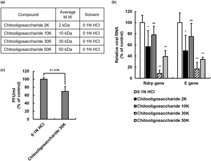
Aims: The discovery of antiviral substances to respond to COVID-19 is a global issue, including the field of drug development based on natural materials. Here, we showed that chitosan-based substances have natural antiviral properties against SARS-CoV-2 in vitro.

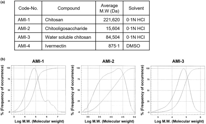
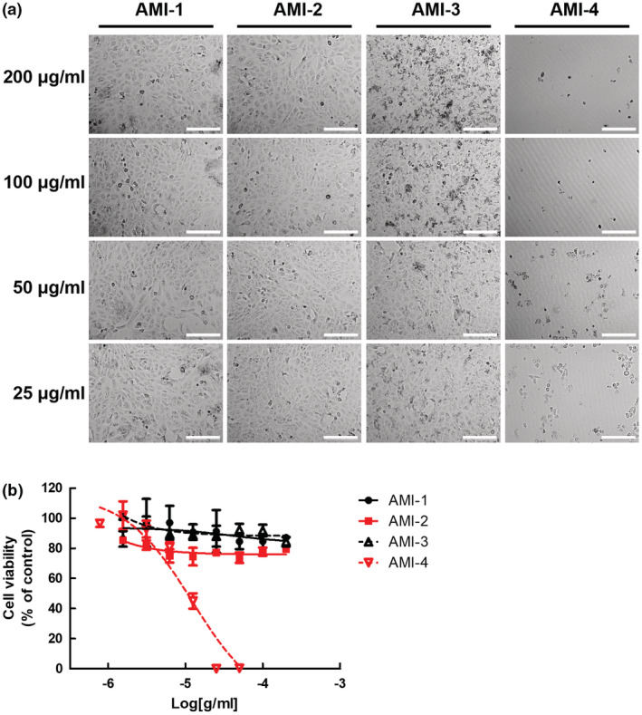
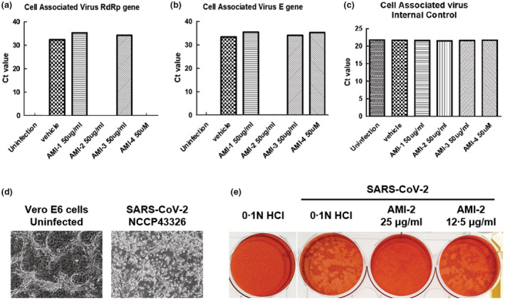
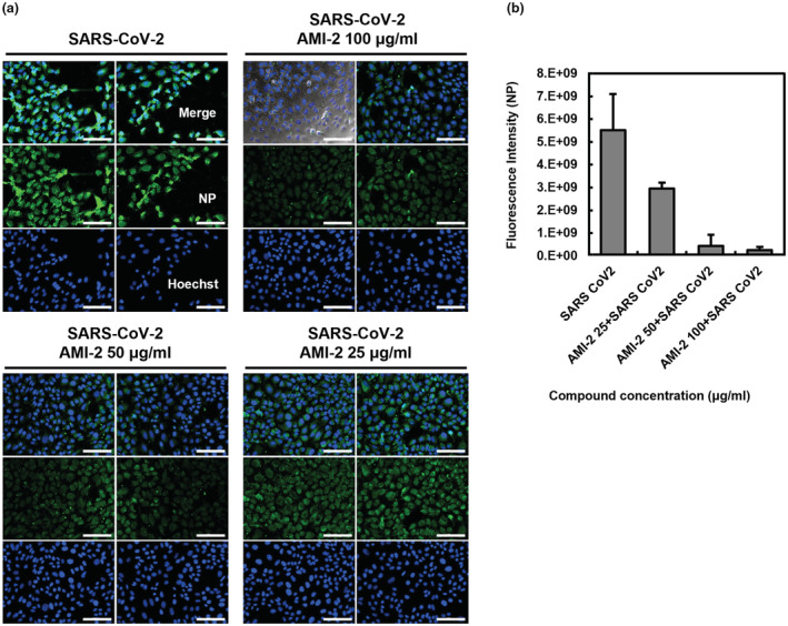
Methods and results: The molecular weight of chitosan-based substances was measured by the gel permeation chromatography analysis. In MTT assay, the chitosan-based substances have low cytotoxicity to Vero cells. The antiviral effect of these substances was confirmed by quantitative viral RNA targeting the RdRp and E genes and plaque assay. Among the substances tested, low molecular weight chitooligosaccharide decreased the fluorescence intensity of SARS-CoV-2 nucleocapsid protein of the virus-infected cells in a dose-dependent manner.

Conclusions: In conclusion, the chitooligosaccharide, a candidate for natural treatment, has antiviral effects against the SARS-CoV-2 virus in vitro.

Significance and impact of study: In this study, it was suggested for the first time that chitosan-based substances such as chitooligosaccharide can have an antiviral effect on SARS-CoV-2 in vitro.

Keywords: COVID-19; SARS-CoV-2; antiviral effect; chitooligosaccharide; natural treatment.


Conflict of interest statement

The authors declare that there are no conflicts of interest. Through Amicogen (Republic of Korea), the authors have filed patent applications on the chitosan‐derivates substances. The authors declare that the research was conducted without any commercial or financial relationships that could be construed as a potential conflict of interest.

Figures

FIGURE 1
FIGURE 1
The average molecular weight of chitosan‐based substances. (a) The candidate substances were used for antiviral effects to SARS‐CoV‐2 in the experiment. Synthetic substances such as chitosan‐based substances and ivermectin are named as follows; AMI‐1 (chitosan), AMI‐2 (chitooligosaccharide), AMI‐3 (water soluble chitosan) and AMI‐4 (ivermectin). (b) Measuring the molecular weight of chitosan‐based substances through gel permeation chromatography. The molecular weight of the chitosan‐based substances of 5% concentration was analysed by chromatography and compared with the standard curve of the standard product.
FIGURE 2
FIGURE 2
Cytotoxicity of chitosan‐based substances on cultured cells. (a) Morphology of chitosan‐based substances and ivermectin treated cells. Phase‐contrast microscopy images were obtained from Vero E6 cells treated with indicated concentrations (25–200 μg/ml) of AMI‐1 to −4 for 24 h. Representative single optical sections are shown. Scale bars, 200 μm. (b) Measurement of cytotoxicity of cells treated with chitosan‐based substances and ivermectin. MTT assay of Vero E6 cells treated with indicated concentration (25–200 μg/ml) of AMI‐1 to −4 for 24 h. Data are represented as mean ± SD.
FIGURE 3
FIGURE 3
Effects of chitosan‐based substances on SARS‐CoV‐2. (a–c) Antiviral effect of chitosan‐based substances on SARS‐CoV‐2 through qPCR analysis. The amount of SARS‐CoV‐2 virus at MOI 0.01 was incubated with 50 μg/ml of chitosan‐based substances (AMI‐1, −2, and −3) or 50 μM of AMI‐4 for 1 h at room temperature, infected with Vero E6 cells, and incubated with the substances for 48 h. Viral RNA was extracted from the culture medium of the cells, and the SARS‐CoV‐2 gene was detected through qPCR analysis. The graph of Ct value was represented to (a) RNA‐dependent RNA polymerase (RdRP) gene, (b) envelope (E) gene of SARS‐CoV‐2 and (c) internal control of qPCR kits, respectively. (d) The cytopathic effect of SARS‐CoV‐2 on Vero E6 cells. The images of cytopathic effects of Vero E6 cells infected with SARS‐CoV‐2 (NCCP43326) for 4 days were obtained by phase‐contrast microscopy images. Representative single optical sections are shown. Scale bars, 100 μm. (e) Plaque assay of SARS‐CoV‐2 infected Vero E6 cells. After 1 h of the virus within indicated concentration of AMI‐2 adsorption, low‐melting agar containing first overlays were added. Secondary overlays were added after 5 days, and the cells were incubated overnight. Use a white‐light transilluminator (light box) to aid in visualize the plaques.
FIGURE 4
FIGURE 4
Dose‐dependent antiviral effect of chitooligosaccharide on SARS‐CoV‐2 infected Vero cells. (a, b) Indirect immunofluorescence images of nucleocapsid protein (NP) in SARS‐CoV‐2 infected Vero E6 cells. Vero E6 cells were infected with SARS‐CoV‐2 at MOI 0.01 within indicated concentrations of the AMI‐2 for 36 h. The infected cells were fixed and stained with anti‐nucleocapsid protein antibody followed by Alexa 488‐labelled antibody and analysed by fluorescence microscopy. The nuclei were stained with Hoechst dye. (a) Representative two different single optical sections are shown. Scale bars, 100 μm. (b) The bar graph was converted from the fluorescence intensity of the images. Data are represented as mean ± SD.
FIGURE 5
FIGURE 5
Antiviral effect of chitooligosaccharides of various molecular weights on SARS‐CoV‐2. (a) The chitooligosaccharides of various molecular weights. Separation by molecular weight to confirm the inhibitory effect of chitooligosaccharide on SARS‐CoV‐2 virus replication. (b) Antiviral effect of chitooligosaccharides on SARS‐CoV‐2 through qPCR analysis. The amount of SARS‐CoV‐2 virus at MOI 0.01 was incubated with 25 μg/ml of chitooligosaccharides (2, 10, 30 and 50 kDa) for 1 h at room temperature, infected with Vero E6 cells, and incubated with the chitooligosaccharides for 48 h. Viral RNA was extracted from the culture medium of the cells, and the SARS‐CoV‐2 gene was detected through qPCR analysis. The graph of relative viral RNA was represented to RdRP gene and E gene of SARS‐CoV‐2. Data are represented as mean ± SD (n = 5, ns. not significant, *p < 0.05, **p < 0.001, two‐way ANOVA). (c) Plaque assay of SARS‐CoV‐2 infected Vero E6 cells. After 1 h of the virus within 200 μg/ml of chitooligosaccharide 30 kDa adsorption, low‐melting agar containing overlays were added. After 3 days, the cells were fixed and stained with crystal violet. The plaque forming unit is calculated by plaque number counting. Data are represented as mean ± SD (n = 3, p < 0.05, Student's t test).

References

    1. Artan, M. , Karadeniz, F. , Karagozlu, M.Z. , Kim, M.M. & Kim, S.K. (2010) Anti‐HIV‐1 activity of low molecular weight sulfated chitooligosaccharides. Carbohydrate Research, 345, 656–662. - PubMed
    1. Bello, M. (2021) Elucidation of the inhibitory activity of ivermectin with host nuclear importin α and several SARS‐CoV‐2 targets. Journal of Biomolecular Structure & Dynamics, 10, 1–9. - PMC - PubMed
    1. Caly, L. , Druce, J.D. , Catton, M.G. , Jans, D.A. & Wagstaff, K.M. (2020) The FDA‐approved drug ivermectin inhibits the replication of SARS‐CoV‐2 in vitro. Antiviral Research, 178, 104787. - PMC - PubMed
    1. Chatterjee, S.K. , Saha, S. & Munoz, M.N.M. (2020) Molecular pathogenesis, immunopathogenesis and novel therapeutic strategy against COVID‐19. Frontiers in Molecular Biosciences, 7, 196. - PMC - PubMed
    1. Cheung, R.C. , Ng, T.B. , Wong, J.H. & Chan, W.Y. (2015) Chitosan: an update on potential biomedical and pharmaceutical applications. Marine Drugs, 13, 5156–5186. - PMC - PubMed