. 2022 Aug 1;189(4):2281-2297.
doi: 10.1093/plphys/kiac215.

A PLETHORA/PIN-FORMED/auxin network mediates prehaustorium formation in the parasitic plant Striga hermonthica

Affiliations

A PLETHORA/PIN-FORMED/auxin network mediates prehaustorium formation in the parasitic plant Striga hermonthica

Ting Ting Xiao et al. Plant Physiol. .

Abstract

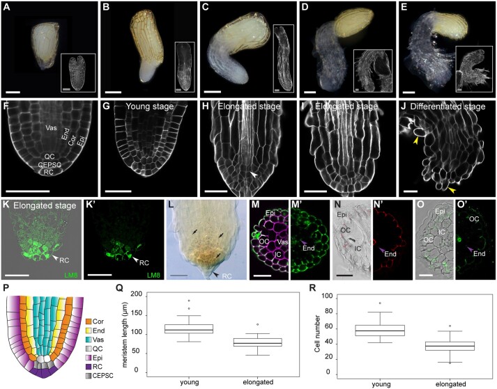
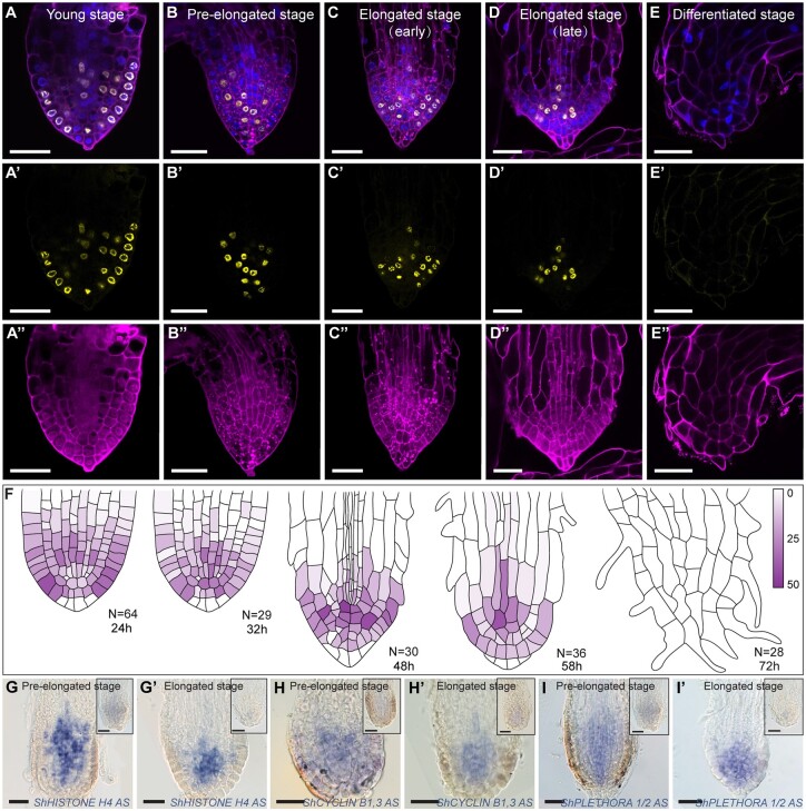
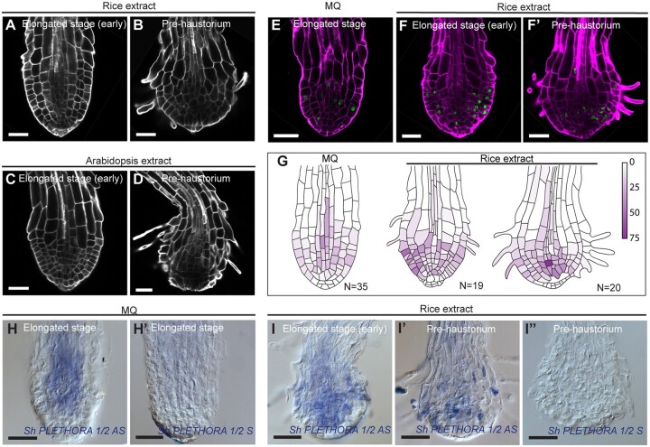
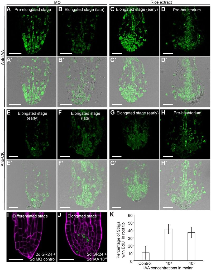
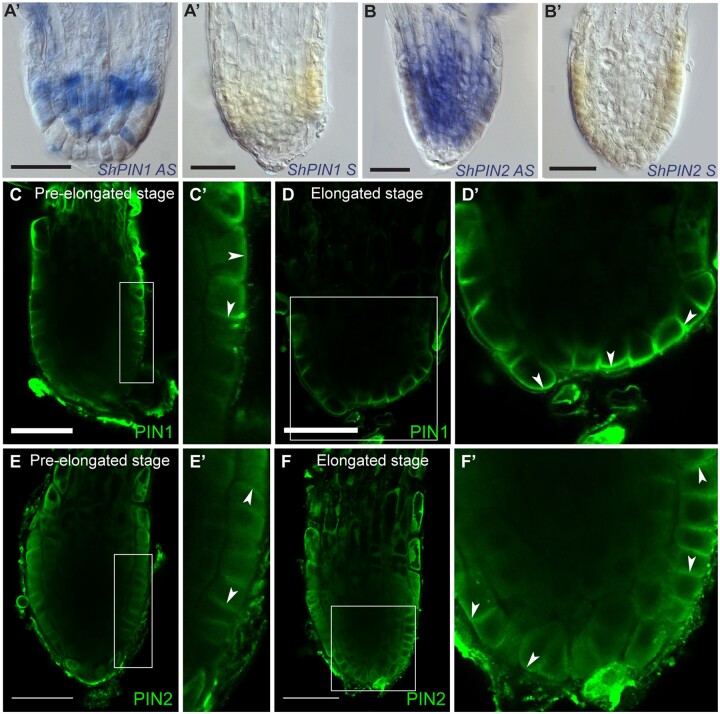
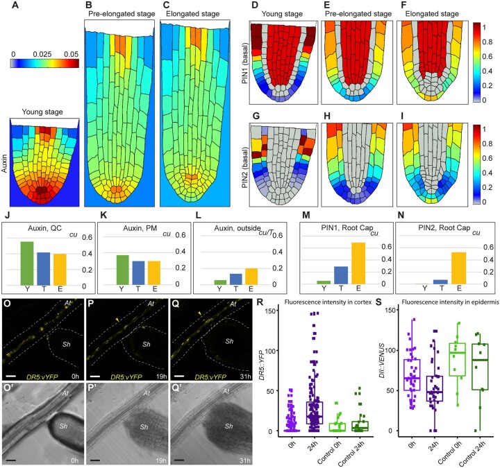
The parasitic plant Striga (Striga hermonthica) invades the host root through the formation of a haustorium and has detrimental impacts on cereal crops. The haustorium results from the prehaustorium, which is derived directly from the differentiation of the Striga radicle. The molecular mechanisms leading to radicle differentiation shortly after germination remain unclear. In this study, we determined the developmental programs that regulate terminal prehaustorium formation in S. hermonthica at cellular resolution. We showed that shortly after germination, cells in the root meristem undergo multiplanar divisions. During growth, the meristematic activity declines and associates with reduced expression of the stem cell regulator PLETHORA1 and the cell cycle genes CYCLINB1 and HISTONE H4. We also observed a basal localization of the PIN-FORMED (PIN) proteins and a decrease in auxin levels in the meristem. Using the structural layout of the root meristem and the polarity of outer-membrane PIN proteins, we constructed a mathematical model of auxin transport that explains the auxin distribution patterns observed during S. hermonthica root growth. Our results reveal a fundamental molecular and cellular framework governing the switch of S. hermonthica roots to form the invasive prehaustoria.


Figures

Figure 1
Figure 1
Cellular organization of the S. hermonthica root meristem. A–E, Macrophotographs of S. hermonthica seedlings at the following different developmental stages: A, seed; B, young; C, pre-elongated; D, elongated; and E, differentiated/prehaustorium. White rectangles show their respective confocal sections stained with Renaissance SCRI stain. F–J, Longitudinal confocal sections of the S. hermonthica roots stained with Renaissance SCRI stain. K–K′, Immunolocalization using the LM8 antibody to mark the root cap in S. hermonthica. (K a confocal overlay of transmission and fluorescence image; K′ is the fluorescent channel). L, Lugol’s iodine stain showing stem cell differentiation. M–O′, Images of Striga root cross sections depicting tissue types using cell wall stains. Sections were taken from above the meristem (150–200 µm distance from the root tip). M, Renaissance stain. M′, Auto-fluorescence. N and N′, Basic fuchsine. O and O′, Berberine stain. P, Schematic representation of the S. hermonthica root meristem at the young stage. Times indicated in the panels are hours after GR24 treatment. Scale bars: macrophotographs in (A–E): 200 µm; Images in (A–E insets; F–O′): 50 µm. Epi, epidermis; OC, outer cortex; Exo, exodermis; IC, inner cortex, End, endodermis; Vas, vasculature; RC, root cap; Cor, cortex; QC, quiescent center (QC); CEPSC, columella and epidermal precursor cells; arrows heads pointing to root hairs (J), root cap (K–L) and casparian strips (M–O′). Q and R are boxplots for meristem size (Q) and cell number (R) in young (N = 86) and elongated (N = 55) S. hermonthica seedlings (24- and 48-h GR24 treatment). N represents the number of seedlings analyzed. Horizontal lines indicate the medians. Box limits indicate the 25th and 75th percentiles. Whiskers extend to the 5th and 95th percentiles. The circles represent outliers.
Figure 2
Figure 2
Prehaustorium formation associates with meristem differentiation. A–E″, Confocal images of S. hermonthica roots stained with the cell wall stain mPS-PI (A″–E″, purple), EdU (A′–E′; yellow), and the nuclear stain Hoechst (blue in A–E). A–A″, young stage; B–B″, pre-elongated stage, C–C″, early elongated stage, D–D″, late elongated stage; E–E″, differentiated stage, F, quantification of the cell division rate during root differentiation. Colored bar indicates the percentage of cell division. RNA in situ hybridization during meristem differentiation in longitudinal sections using ShHistone-H4 (G–G′), ShCYCLIN B1,3 (H–H′), and ShPLETHORA1/2 (I–I′) probes. Roots in the upper right of the images represent the sense control for each probe. Striga hermonthica seedlings with 24- or 48-h GR24 treatment. All scale bars: 50 µm.
Figure 3
Figure 3
Prehaustorium of S. hermonthica induced by host and nonhost root extract treatment. A–D, confocal images of Striga roots treated with rice (A and B) and Arabidopsis (C and D) roots extract. Roots were stained using Renaissance SCRI stain. Roots were treated after 24 h of germination in presence of GR24 and imaged after 4 and 12 h after treatment with root extracts. E and F′, EdU staining showing cell division in the differentiating S. hermonthica roots. G, quantification of the cell division rate during prehaustorium formation. Colored bar indicates the percentage of cell division. H–I″, RNA in situ hybridization during prehaustorium formation in longitudinal sections using ShPLETHORA1/2. H–H′ are roots germinated in MQ water. I–I″ roots treated with rice extract for 12 h after 24 h of germination. MQ, Milli-Q water, AS, Antisense, S, sense control. All scale bars: 50 µm.
Figure 4
Figure 4
Auxin–cytokinin homeostasis control of S. hermonthica meristem activity. A–D′, immunolocalization showing auxin distribution in S. hermonthica root sections using anti-IAA antibody, N = 30; E–H′, cytokinin accumulation using anti-cytokinin antibody. Seedlings are 24 (A and E) and 48 h (B and F) after GR24 treatment-induced germination and 12 h (C, G, D, and H) of rice extract treatment. N = 20; A–D and E–H are fluorescence channel; A′–D′ and E′–H′ are overlay fluorescence and transmission light channels. I–K, IAA application delayed meristem differentiation in the S. hermonthica radicle, as monitored by EdU staining. I and J, Confocal images of control (I) and IAA-treated roots (J). MQ, Milli-Q water. K, Percentage of root containing cells undergoing cell division (EdU-stained cells) in S. hermonthica roots treated with different molar IAA concentrations. Columns indicate the means generated from three biological replicates; error bars represent se. A significant difference from the value of the non-treated roots versus IAA-treated roots was determined by t test (P < 0.05). The number of S. hermonthica seedlings used were as follows for the control (N = 44, 53, 40); 10−8 (N = 43, 45, 48); 10−7 (N = 93, 107, 95). N represents the number of seedlings used from three independent biological replicates. All scale bars: 50 µm.
Figure 5
Figure 5
PIN protein expression and polarity in S. hermonthica during root differentiation. A–B′, RNA in situ hybridization of S. hermonthica root meristems in longitudinal sections using ShPIN1 (A–A′) and ShPIN2 (B–B′). A and B are the antisense probes and A′ and B′ are the sense control probes. C–F′, Immunolocalization using Arabidopsis thaliana PIN1 and PIN2 antibodies showing (C–D′) basal PIN1 in the epidermis. C′ and D′ are insets from C and D, respectively. N = 30. E and F′, PIN2 localization showing epidermal apical, lateral, and basal polarity. Arrows indicate the shift in PIN polarity. E′ and F′ are insets from E and F, respectively. Striga hermonthica seedlings were treated with GR24 for 24 or 48 h. N = 60. All scale bars: 50 µm.
Figure 6
Figure 6
Mathematical model depicting auxin distribution in S. hermonthica during root growth. (A–C), Quasi-steady state in auxin distribution inside and outside the Striga root at three consequent developmental layouts. (A, D, and G) Young stage (Y), (B, E, and H) pre-elongated stage (T), and (C, F, and I) elongated stage (E). D–F, PIN1 localization, G–I, PIN2 localization with basal polarity. PIN2 with apical polarity is depicted in Supplemental Figure S9. The color scale bars are concentration units (cu). J–N, Comparison of the variable values between the stages. Auxin levels in the QC (J); in the proximal meristem (PM) (K); excreted from S. hermonthica roots (L) during calculation time T. M, PIN1 levels in the root cap; N, the level of basally polarized PIN2 in the root cap. cu, concentration units. O–Q′, time-lapse images of germinating S. hermonthica in contact with Arabidopsis roots containing DR5::vYFP. S. hermonthica were used 24 h after germination, N = 10. O–Q, Fluorescence YFP channel. O′–Q′ PMT transmission channel. Arrowheads indicate ectopic DR5::vYFP expression in P, Q at A. thaliana (At). sh, S. hermonthica. Scale bars: 50 µm. R, the DR5 fluorescence values are significantly increased after 24 h when the Striga root differentiates in contact with Arabidopsis roots (two-tailed t test, P  = 0.03794). The DR5 values are not significantly different when physical contact is not established with the Arabidopsis roots (two-tailed t test, P  = 0.4947). S, the DII fluorescence values are significantly decreased after 24 h when the Striga root differentiates in contact with Arabidopsis roots (two-tailed t test, P <2.2e–16) the DII fluorescence are not significantly different when physical contact is not established (two-tailed t test, P  = 0.3095). Control are striga plants that have not been in contact with Arabidopsis. Box limits indicate the 25th and 75th percentiles. Whiskers extend to the 5th and 95th percentiles. Points indicates the data.

References

    1. Abas L, Kolb M, Stadlmann J, Janacek DP, Lukic K, Schwechheimer C, A.Sazanov L, Mach L, Friml J, Hammes UZ (2021) Naphthylphthalamic acid associates with and inhibits PIN auxin transporters. Proc Natl Acad Sci USA 118: e2020857118 - PMC - PubMed
    1. Aida M, Beis D, Heidstra R, Willemsen V, Blilou I, Galinha C, Nussaume L, Noh YS, Amasino R, Scheres B (2004) The P LET HORA genes mediate patterning of the Arabidopsis root stem cell niche. Cell 9: 109–120 - PubMed
    1. Al-Babili S, Bouwmeester HJ (2015) Strigolactones, a novel plant hormone. Annu Rev Plant Biol 66: 161–186 - PubMed
    1. Albrecht H, Yoder JI, Phillips DA (1999) Flavonoids promote haustoria formation in the root parasite Triphysaria versicolor. Plant Physiol 119: 585–591 - PMC - PubMed
    1. Baird W V., Riopel JL (1983) Experimental studies of the attachment of the parasitic angiosperm Agalinis purpurea to a host. Protoplasma 118: 206–218

Publication types

Substances