Review
. 2023 Feb;96(1142):20220281.
doi: 10.1259/bjr.20220281. Epub 2022 May 20.

Imaging incidental adrenal lesions

Affiliations
Review

Imaging incidental adrenal lesions

Anju Sahdev. Br J Radiol. 2023 Feb.

Abstract

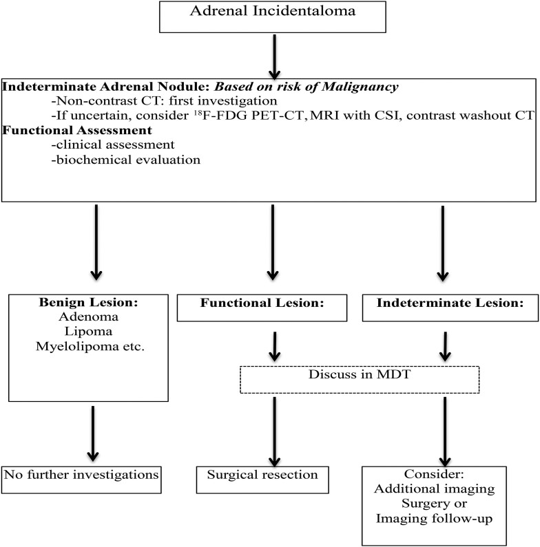
Incidental adrenal masses are among the most common incidental lesions detected on cross-sectional imaging. The majority are benign lesions, adenomas and myelolipomas being the most common. Simple cross-sectional imaging techniques using CT and MRI permit the characterization of over 80%, thereby requiring no further imaging. The remaining lesions are considered indeterminate. These lesions consist of benign and malignant lesions sharing imaging features. Further imaging and management of these indeterminate lesions should be guided by close collaboration between different specialists in an MDT setting. Advanced imaging options include dedicated adrenal scintigraphy, positron emission tomography CT, biopsy and surveillance. Biochemical and hormonal evaluation is also important to identify hyperfunctioning adrenal lesions. This review focuses on imaging features of benign and malignant adrenal masses used for characterization and suggests an imaging pathway for indeterminate adrenal masses.


Figures

Figure 1.
Figure 1.
Bilateral adrenal incidental lesions. Adrenal washout protocol CT. (A) Non-contrast enhanced CT. (B) Post-contrast enhanced CT at 60 s. (C) Post- contrast-enhanced CT at 15 min. Left adrenal nodule Pre C = 13 HU, PV C = 68 HU, Delayed C = 22 HU. Absolute washout = 46/55 = 84% in keeping with lipid poor adenoma. Right nodule Pre C = 21 HU, PV C = 64 HU, delayed C = 25 HU. Absolute washout = 39/43 = 91% also in keeping with a lipid poor adenoma. HU, Hounsfield unit.
Figure 2.
Figure 2.
Lipid rich adenoma on MRI. (A) Axial T 2 weighted image showing an incidentally detected left adrenal mass with typically low T2 signal intensity (arrow). (B) In-phase axial chemical shift imaging showing the lesion with intermediate signal intensity value 948. (C) Out-of-phase axial chemical shift image showing a visual loss of signal intensity value 219. This provides a SII drop of 77% in keeping with an adenoma. (D) NCCT showing a left adrenal mass with a CT attenuation of HU-3HU confirming a benign lipid rich adenoma. NCCT, non-contrast CT.
Figure 3.
Figure 3.
Adrenal cyst. (A) Single phase portovenous CT with a left adrenal mass 18 HU in density (arrow). (B) Axial in-phase T1 and out-of-phase weighted images with the lesion demonstrating a uniform low fluid signal intensity and no signal loss. C: Axial T2 weighed image: showing high fluid signal in keeping with a simple cyst. This has remained stable over a 5 year follow-up on imaging performed for unrelated indications. HU, Hounsfield unit.
Figure 4.
Figure 4.
Features of myelolipomas. (A) Axial T 1 weighted image. (B) Axial T 1 weighted image with fat saturation. A large right bilobed mass with signal similar to the retroperitoneal fat on all sequences. The anterior lobule is fatty and the posterior lobule is an admixture of fat and myeloid components (arrows). (C) Out-of-phase chemical shift image showing no loss of signal in the pure fatty anterior lobule and limited signal loss (broken arrow) in the admixed tissue in keeping with a myelolipoma. (D) Non-contrast CT. (E) Post-contrast CT acquired 60 s following i.v. contrast injection A 3 cm mass is demonstrated in the left adrenal gland (arrows). The mass is mainly composed of homogenous solid enhancing tissue. Small foci of fat are seen scattered within the mass (dashed arrows). The mass was stable over 4 years with no functional activity. These features are consistent with a fat poor myelolipoma.
Figure 5.
Figure 5.
Features of pheochromocytomas. (A) Post-contrast CT acquired 60 s following i.v. contrast injection demonstrating a large heterogenous mass in the right adrenal with multiple cystic areas. (B) Portovenous phase CT of the abdomen showing heterogeneity in the mass with solid areas in the rim. (C) Axial T 2 weighted image with a high T2 signal intensity mass in the left adrenal (arrows). (D) Axial T1 in-phase and out-of-phase images with the mass demonstrating no loss of signal intensity. (E) Post-gadolinium-enhanced images showing marked enhancement typical for a pheochromocytoma.
Figure 6.
Figure 6.
Adrenal abscess in acute TB. Coronal reconstructed post-contrast CT in a patient presenting with scrotal cutaneous abscesses. On CT, there are bilateral adrenal low-density masses with internal septation and enhancement (arrows), left renal pelvis urothelial thickening (dashed arrows) and a right pelvic filling defect in keeping with TB. This was confirmed on drainage of the scrotal abscess and all sites of disease resolved following tuberculous treatment. TB, tuberculosis.
Figure 7.
Figure 7.
Adrenal hemorrhage in a trauma patient. (A) NCCT demonstrating high density irregular ‘mass’ measuring 34 HU (arrows). (B) CECT coronal reconstruction showing no increase in density in keeping with a retroperitoneal adrenal hemorrhage. (C) Spectral CT iodine map image shows no iodine uptake within the adrenal bed, excluding an underling mass lesion. (D) 3 month follow-up axial T 1 weighted MRI image showing a significant reduction in size of the hemorrhage and only a small residual hematoma. CECT, contrast-enhanced CT; HU, Hounsfield unit; NCCT, non-contrast CT.
Figure 8.
Figure 8.
68Ga Somatostatin Receptor PET-CT for metastatic pheochromocytoma. (A) Coronal T 2 weighted image with a large right- sided adrenal malignant heterogenous mass encircling and invading the IVC (arrow). Associated retroperitoneal lymphadenopathy is present (dashed arrow). (B) Coronal fused 68Ga PET CT performed for staging and evaluation of metastatic tumor bulk confirms peripheral uptake in the right adrenal mass and distant metastases in the neck, chest and retroperitoneum. IVC, inferior vena cava; PET, positron emission tomography.
Figure 9.
Figure 9.
Atypical adenoma on 18F-FDG PET CT in a patient with clinical and biochemical Cushing’s syndrome. (A) Non-contrast CT: a right adrenal homogenous mass with a density of 16 HU. (B) Post-contrast CT acquired 60 s following i.v. contrast injection: The mass has a density of 61 HU with no heterogeneity, vascular invasion or distant metastases. (C) Post-contrast CT acquired 15 min following i.v. contrast injection: contrast washout is observed and the density is 31 HU. The mass therefore has an absolute contrast washout of 68% and relative washout of 51% in keeping with a lipid poor adenoma. (D) Axial fused 18F-FDG PET CT image of the adenoma demonstrating marked 18F-FDG avidity with an SUV max of 6.9. This raises the concern for an ACC; however, on histology, it was a benign functioning adenoma only with no suspicious or borderline features to suggest a ACC. ACC, adrenocortical carcinomas; FDG, fludeoxyglucose; HU, Hounsfield unit; PET, positron emission tomography; SUV, standardized uptake value.
Figure 10.
Figure 10.
Cystic metastases from colorectal adenocarcinoma. Coronal reconstruction of single portovenous phase CT. A heterogeneous right adrenal mass is seen with central necrosis and ill-defined margins suggestive of a malignant lesion (arrows). An associated large hepatic flexure colonic carcinoma is also present (dashed arrows). Post-treatment response with chemotherapy was seen in both lesions and final histology confirmed an adrenal metastases.
Figure 11.
Figure 11.
Malignant adrenocortical carcinomas. (A) PV phase CT showing a left adrenal incidentaloma with a post-contrast measurement of 61 HU (arrow). (B) PV phase adrenal CT 10 months later: the indeterminate nodule was not fully characterized at presentation but followed-up. The lesion has significantly increased in size and demonstrates malignant features of diaphragmatic invasion (dashed arrow). The lesion had a pre-contrast density of 39 HU and delayed density of 52 HU (not shown) providing an absolute contrast washout of 39%. Biopsy confirmed an adrenocortical carcinoma. (C) Adrenocortical carcinoma with. Weiss score 7 (aggressive/malignant behavior) ( T)umor thrombus invading the adrenal and renal vein extending to the IVC (dashed arrows). (D) Low ADC of the mass and tumor vascular thrombus (dashed arrows). ADC, apparent diffusion coefficient; IVC, inferior vena cava; HU, Hounsfield unit; PV, portovenous.
Figure 12.
Figure 12.
Biopsy confirmed adrenal Hodgkin’s lymphoma. (A) Single post- contrast portovenous phase CT: the mass demonstrates poor enhancement and no heterogeneity despite the large size. Several thoracic enlarged nodes were also present (not demonstrated). The differential of this large homogenous mass includes lymphoma or a benign neurogenic tumor. In the presence of distant lymphadenopathy, lymphoma is more likely. (B) Coronal reconstructed fused 18F-FDG PET CT: the mass demonstrates marked avidity with an SUV max of 7. This again is more typical of lymphoma as benign neurogenic tumors have low grade avidity. FDG, fludeoxyglucose; HU, Hounsfield unit; PET, positron emission tomography; SUV, standardized uptake value.
Figure 13.
Figure 13.
Flowchart on the management of patients with adrenal incidentaloma. Based on ENSAT guideline on adrenal incidentalomas. ENSAT, European Society of Endocrinology and European Network for the Study of Adrenal Tumors.

Similar articles

Cited by

References

    1. Barzon L, Sonino N, Fallo F, Palu G, Boscaro M. Prevalence and natural history of adrenal incidentalomas. Eur J Endocrinol 2003; 149: 273–85. doi: 10.1530/eje.0.1490273 - DOI - PubMed
    1. Song JH, Chaudhry FS, Mayo-Smith WW. The incidental adrenal mass on CT: prevalence of adrenal disease in 1,049 consecutive adrenal masses in patients with no known malignancy. AJR Am J Roentgenol 2008; 190: 1163–68. doi: 10.2214/AJR.07.2799 - DOI - PubMed
    1. Frilling A, Tecklenborg K, Weber F, Kühl H, Müller S, Stamatis G, et al. . Importance of adrenal incidentaloma in patients with a history of malignancy. Surgery 2004; 136: 1289–96. doi: 10.1016/j.surg.2004.06.060 - DOI - PubMed
    1. Oliver TW Jr, Bernardino ME, Miller JI, Mansour K, Greene D, Davis WA. Isolated adrenal masses in nonsmall-cell bronchogenic carcinoma. Radiology 1984; 153: 217–18. doi: 10.1148/radiology.153.1.6473783 - DOI - PubMed
    1. Iuga AC, Marboe CC, M Yilmaz M, Lefkowitch JH, Gauran C, Lagana SM. Adrenal vascular changes in COVID-19 autopsies. Arch Pathol Lab Med 2020; 144: 1159–60. doi: 10.5858/arpa.2020-0248-LE - DOI - PubMed