. 2022 Jul 5;50(W1):W90-W98.
doi: 10.1093/nar/gkac345.

BeStSel: webserver for secondary structure and fold prediction for protein CD spectroscopy

Affiliations

BeStSel: webserver for secondary structure and fold prediction for protein CD spectroscopy

András Micsonai et al. Nucleic Acids Res. .

Abstract

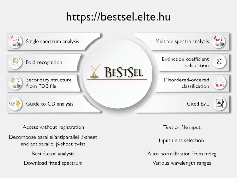
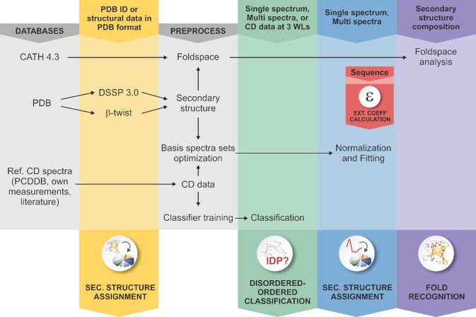
Circular dichroism (CD) spectroscopy is widely used to characterize the secondary structure composition of proteins. To derive accurate and detailed structural information from the CD spectra, we have developed the Beta Structure Selection (BeStSel) method (PNAS, 112, E3095), which can handle the spectral diversity of β-structured proteins. The BeStSel webserver provides this method with useful accessories to the community with the main goal to analyze single or multiple protein CD spectra. Uniquely, BeStSel provides information on eight secondary structure components including parallel β-structure and antiparallel β-sheets with three different groups of twist. It overperforms any available method in accuracy and information content, moreover, it is capable of predicting the protein fold down to the topology/homology level of the CATH classification. A new module of the webserver helps to distinguish intrinsically disordered proteins by their CD spectrum. Secondary structure calculation for uploaded PDB files will help the experimental verification of protein MD and in silico modelling using CD spectroscopy. The server also calculates extinction coefficients from the primary sequence for CD users to determine the accurate protein concentrations which is a prerequisite for reliable secondary structure determination. The BeStSel server can be freely accessed at https://bestsel.elte.hu.


Figures

Graphical Abstract
Graphical Abstract
Functions of the BeStSel web server for the analysis of protein CD spectra.
Figure 1.
Figure 1.
Schematic representation of the BeStSel server. Block diagram shows the modules and function of the BeStSel package. Arrows indicate the input and output data. From a single CD spectrum the secondary structure contents are estimated and then, based on these, the protein fold can be predicted. A series of CD spectra as input can be evaluated at once to get the secondary structure contents. Users can provide arbitrary secondary structure contents and carry out the fold prediction for that secondary structure composition. Users can also enter PDB IDs or upload structure files in PDB format as input to find the corresponding secondary structure contents and fold classification. Based on the CD data, a binary disordered-ordered classification can be carried out. To aid correct concentration determination, extinction coefficients at 205 and 214 nm can be calculated from the primary sequence of the protein (15,16).
Figure 2.
Figure 2.
The effect of environmental conditions on the protein structure studied by CD spectroscopy and analyzed by the BeStSel web server. (A, B) CD spectra of wild type (A) and D76N mutant β2m (B) were recorded at various pH values in 10 mM Na-citrate buffer at 37°C. (C) Secondary structure contents provided by BeStSel were added up as α-helix, β-sheet and turn + others. The mutant protein (red) is more sensitive to pH drop than the wild-type one (blue) and starts to loose its β-structure below pH 6.0. CD measurements were carried out on a benchtop Jasco J-1500 spectropolarimeter (1 mm pathlength, 50 nm/min scan rate, 2 sec response time, 1 nm bandwidth, accumulation: 6). (D) The two β2m variants exhibit very similar high-resolution native structure making difficult to explain the difference in the pathology. (E) Insulin can exhibit various conformations depending on the solution conditions. Its native, monomeric state is α-helical. Under nonnative conditions, such as low pH and the presence of alcohols, it forms β-structured aggregates with different β-sheet compositions as shown in (F). At pH 2.0, it forms amyloid fibrils with characteristic parallel β-sheets. These spectra were collected by SRCD at DISCO beamline in SOLEIL Synchrotron, France.

Similar articles

Cited by

References

    1. Wallace B.A. Synchrotron radiation circular-dichroism spectroscopy as a tool for investigating protein structures. J. Synchrotron Radiat. 2000; 7:289–295. - PubMed
    1. Greenfield N.J. Using circular dichroism spectra to estimate protein secondary structure. Nat. Protoc. 2006; 1:2876–2890. - PMC - PubMed
    1. Khrapunov S. Circular dichroism spectroscopy has intrinsic limitations for protein secondary structure analysis. Anal. Biochem. 2009; 389:174–176. - PMC - PubMed
    1. Micsonai A., Wien F., Kernya L., Lee Y.H., Goto Y., Refregiers M., Kardos J.. Accurate secondary structure prediction and fold recognition for circular dichroism spectroscopy. Proc. Natl. Acad. Sci. U.S.A. 2015; 112:E3095–E3103. - PMC - PubMed
    1. Sillitoe I., Bordin N., Dawson N., Waman V.P., Ashford P., Scholes H.M., Pang C.S.M., Woodridge L., Rauer C., Sen N.et al. .. CATH: increased structural coverage of functional space. Nucleic Acids Res. 2021; 49:D266–D273. - PMC - PubMed

Publication types

Substances