Review
. 2022 Dec;219(6):854-868.
doi: 10.2214/AJR.22.27635. Epub 2022 May 11.

Imaging Surveillance Options for Individuals With a Personal History of Breast Cancer: AJR Expert Panel Narrative Review

Affiliations
Review

Imaging Surveillance Options for Individuals With a Personal History of Breast Cancer: AJR Expert Panel Narrative Review

Marissa B Lawson et al. AJR Am J Roentgenol. 2022 Dec.

Abstract

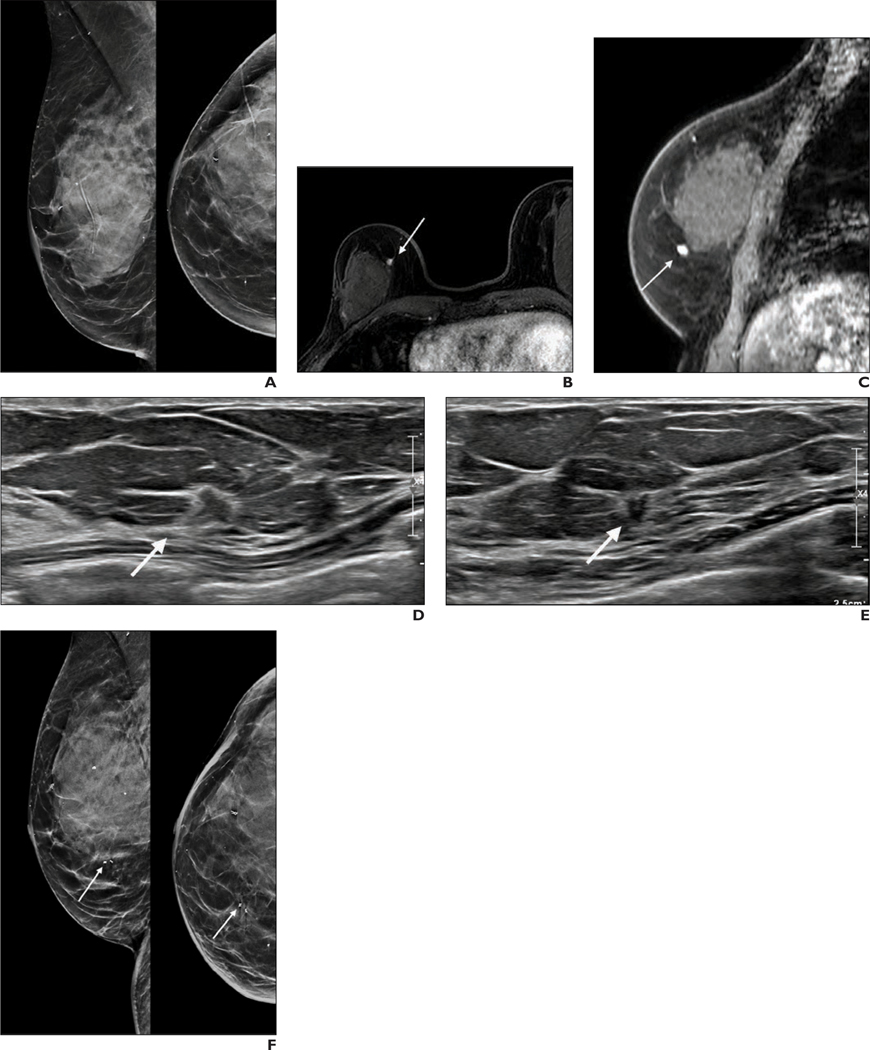
Annual surveillance mammography is recommended for breast cancer survivors on the basis of observational studies and meta-analyses showing reduced breast cancer mortality and improved quality of life. However, breast cancer survivors are at higher risk of subsequent breast cancer and have a fourfold increased risk of interval breast cancers compared with individuals without a personal history of breast cancer. Supplemental surveillance modalities offer increased cancer detection compared with mammography alone, but utilization is variable, and benefits must be balanced with possible harms of false-positive findings. In this review, we describe the current state of mammographic surveillance, summarize evidence for supplemental surveillance in breast cancer survivors, and explore a risk-based approach to selecting surveillance imaging strategies. Further research identifying predictors associated with increased risk of interval second breast cancers and development of validated risk prediction tools may help physicians and patients weigh the benefits and harms of surveillance breast imaging and decide on a personalized approach to surveillance for improved breast cancer outcomes.

Keywords: breast MRI; breast cancer surveillance; contrast-enhanced mammography; mammography; personal history of breast cancer; risk based; supplemental surveillance; whole-breast ultrasound.


Figures

Figure 1.
Figure 1.
Current state of surveillance mammography and outcomes
Figure 2.
Figure 2.
Second breast cancer detected by surveillance mammography. 54-year-old woman with a history of left breast ductal carcinoma in situ (DCIS) treated with lumpectomy and whole breast radiation. (A) Mediolateral oblique (left) and craniocaudal (right) views at surveillance mammography 8 years after treatment show new retroareolar microcalcifications. (B) Magnified mediolateral (left) and craniocaudal (right) views confirm suspicious microcalcifications. Pathology from stereotactic guided biopsy showed multiple foci of microinvasive carcinoma in a background of extensive high-grade DCIS (ER+, PR-, HER2-).
Figure 3.
Figure 3.
Second breast cancer detected by surveillance MRI. 53-year-old woman with a history of right breast invasive ductal carcinoma, ER+, PR+, and HER2-, treated with partial mastectomy, chemotherapy, radiation, and 5 years of tamoxifen. (A) Mediolateral oblique (left) and craniocaudal (right) views at surveillance mammography 10 years after treatment were given a benign assessment. (B) Axial and (C) sagittal contrast-enhanced T1-weighted MR images from a surveillance MRI performed seven months later show a 4 mm irregular enhancing right breast mass with rapid initial and delayed washout kinetic features (arrow). (D) Radial and (E) antiradial views show a sonographic correlate (arrow). (F) Mediolateral oblique (left) and craniocaudal (right) views from a post-biopsy mammogram demonstrate the biopsy marker clips appropriately positioned (arrow). Pathology showed new primary breast cancer (invasive lobular carcinoma; ER+, PR+, HER2-).
Figure 4.
Figure 4.
False-positive finding on surveillance MRI. 36-year-old woman with a history of right breast invasive ductal carcinoma with ductal carcinoma in situ (ER+, PR+, HER2 -), treated with mastectomy and endocrine therapy and who presented for high-risk screening MRI 4 months after surgery. (A) Axial and (B) sagittal contrast-enhanced T1-weighted MR images of the left breast show non-mass enhancement at 1 o’clock, measuring 11 mm and having rapid initial kinetics. The finding was biopsied using MRI guidance (images not shown). (C) Mediolateral oblique (left) and craniocaudal (right) views from post-biopsy mammogram show clip in the expected location (arrow). Pathology results revealed benign stromal fibrosis and focal apocrine metaplasia, concordant with the imaging findings.
Figure 5.
Figure 5.
Second breast cancer detected by surveillance ultrasound. 42-year-old woman with a history of left breast invasive ductal carcinoma (IDC) (ER +, PR+, and HER2-), treated with lumpectomy and radiation. (A) Mediolateral oblique (left) and craniocaudal (right) views from baseline posttreatment mammogram were given a benign assessment. (B) Antiradial image from a surveillance ultrasound performed 6 months later revealed an irregular mass adjacent to the lumpectomy bed measuring 9 × 7 × 6 mm. (C) Mediolateral (left) and craniocaudal (right) views from a post-biopsy mammogram show the biopsy marker clip adjacent to the lumpectomy bed. Pathology was recurrent IDC (grade 2; ER +, PR+, HER2 equivocal).
Figure 6.
Figure 6.
Second breast cancer detected by contrast-enhanced mammography (CEM). 70-year-old woman with a history of left breast invasive ductal carcinoma (IDC) (ER+, PR+, HER2-), treated with lumpectomy, radiation, and 7 years of endocrine therapy. (A) Mediolateral oblique (left) and craniocaudal (right) recombined images from surveillance CEM 11 years after treatment show non-mass enhancement in the upper outer quadrant of the left breast (arrow) (low-energy images not shown). Pathology from MRI-guided biopsy showed in-breast recurrence (IDC and ductal carcinoma in situ; ER+, PR+, HER2-).
Figure 7.
Figure 7.
Breast cancer survivors eligible for supplemental surveillance with MRI. In a cohort of 30,954 women receiving surveillance mammography in U.S. community practice [8], a total of 61.8% (those in the three cells with darker shading and thicker border) would be eligible for supplemental surveillance MRI based on American College of Radiology criteria of age less than 50 years at first breast cancer diagnosis or dense breasts on surveillance mammography.

References

    1. Houssami N, Ciatto S. Mammographic surveillance in women with a personal history of breast cancer: how accurate? How effective? Breast 2010; 19:439–445 - PubMed
    1. Lu WL, Jansen L, Post WJ, Bonnema J, Van de Velde JC, De Bock GH. Impact on survival of early detection of isolated breast recurrences after the primary treatment for breast cancer: a meta-analysis. Breast Cancer Res Treat 2009; 114:403–412 - PubMed
    1. Buist DS, Bosco JL, Silliman RA, et al. Long-term surveillance mammography and mortality in older women with a history of early stage invasive breast cancer. Breast Cancer Res Treat 2013; 142:153–163 - PMC - PubMed
    1. Paszat LF, Sutradhar R, Gu S, Rakovitch E. Annual surveillance mammography after early-stage breast cancer and breast cancer mortality. Curr Oncol 2016; 23:e538–e545 - PMC - PubMed
    1. Buist DS, Abraham LA, Barlow WE, et al. Diagnosis of second breast cancer events after initial diagnosis of early stage breast cancer. Breast Cancer Res Treat 2010; 124:863–873 - PMC - PubMed