

doi: 10.1371/journal.pone.0265247.
eCollection 2022.
Spectral homogeneity of human platelets investigated by SERS
Affiliations
- PMID: 35544536
- PMCID: PMC9094501
- DOI: 10.1371/journal.pone.0265247
Item in Clipboard
Spectral homogeneity of human platelets investigated by SERS
PLoS One.
.
Abstract
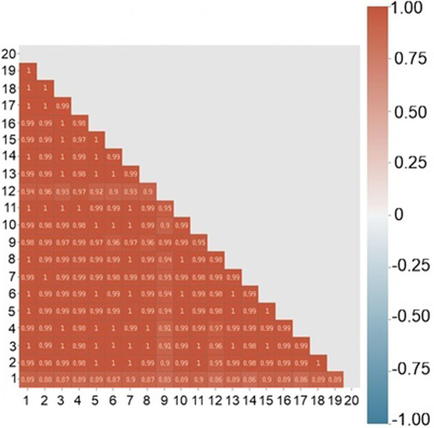
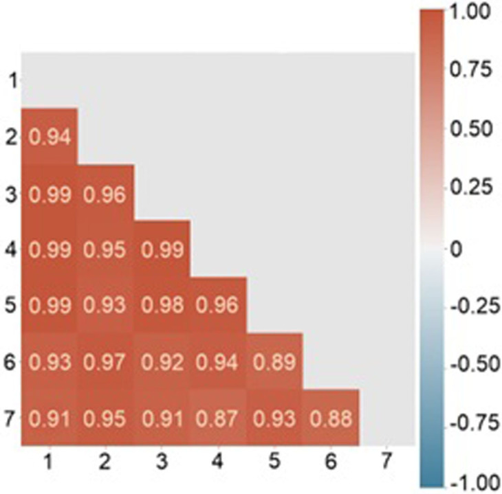
This paper describes a detailed study of the spectral homogeneity of human platelets using Surface-enhanced Raman spectroscopy (SERS). We used a combined approach based on multivariate methods as principal component analysis and pair correlation algorithms to investigate platelets spectral properties. The correlation coefficients for each sample have been calculated, and the average coefficient of determination has been estimated. The high degree of spectral homogeneity inside one probe and between them has been revealed. The prospects of obtained results usage for pathologies based on platelet conformations during cardiovascular diseases have been demonstrated.
Conflict of interest statement
The authors have declared that no competing interests exist.
Figures
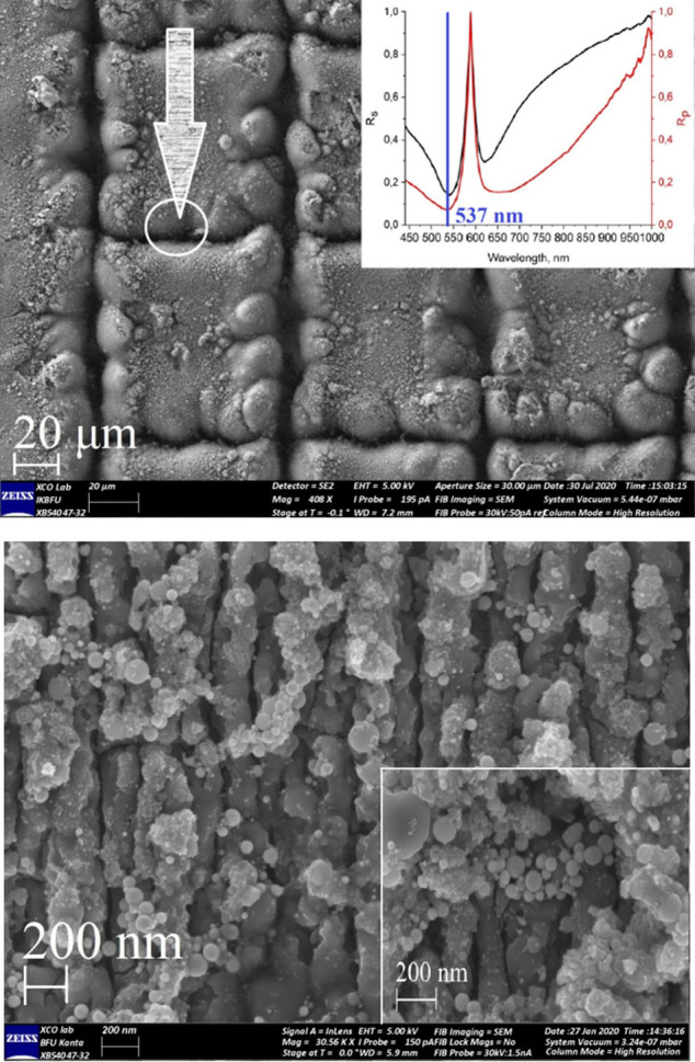
20 μm scale (a) with Au NPs and 1 μm scale (b), 200 nm scale (b, inset) with Au NPs. 1a (inset) illustrates results for spectral ellipsometry experiments for Ti/Au surface.
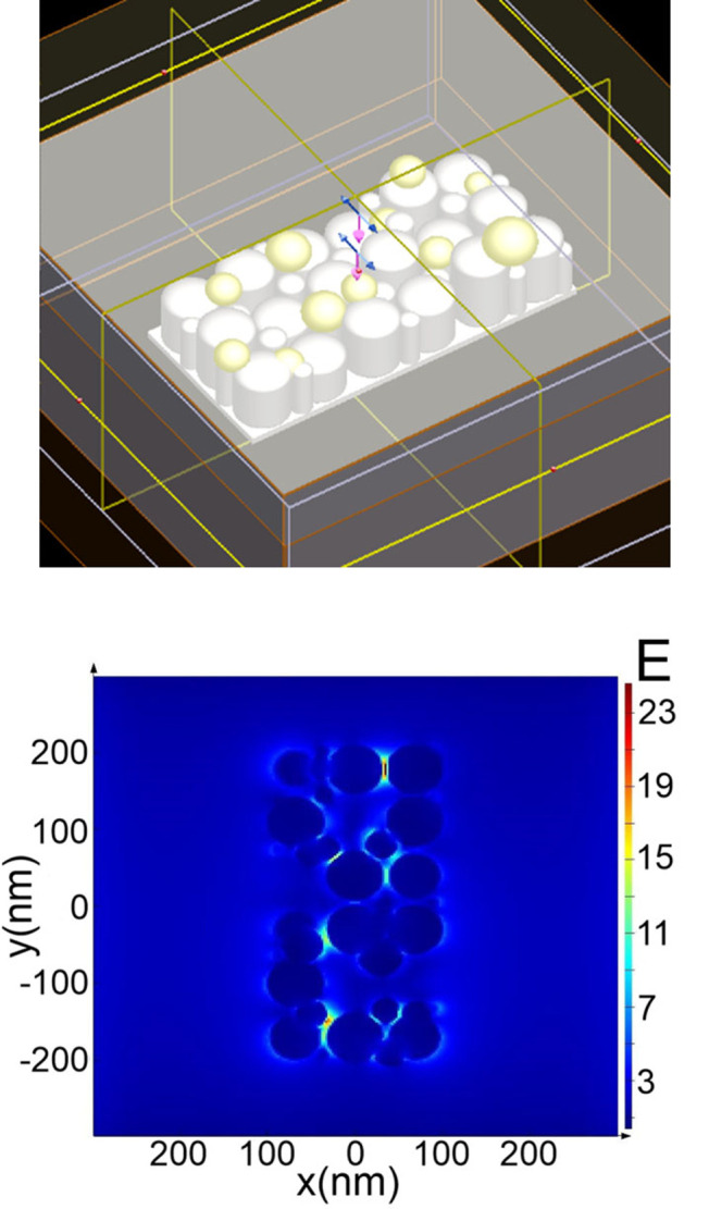
FDTD model area with “grating” surface geometry (a) and electric field strength distribution (b).
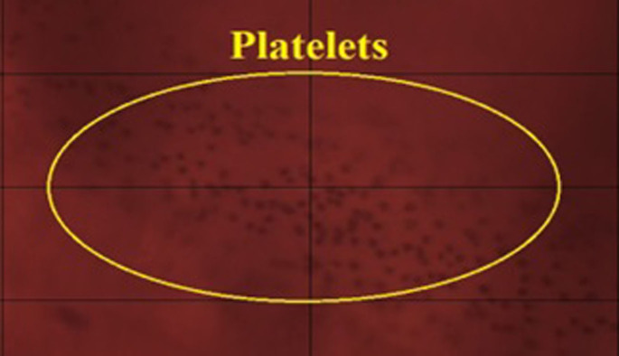
100x optical image of the platelet mass, deposited on Ti/Au surface.
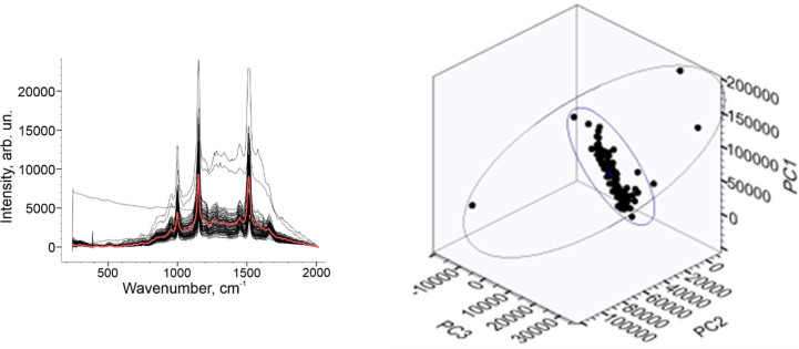
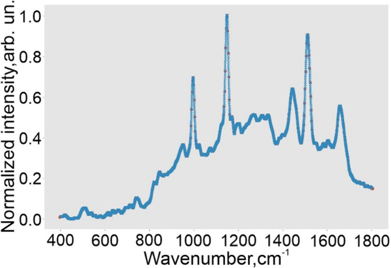
Raman spectral dataset (a) and PCA 3D plot of platelets set (b).
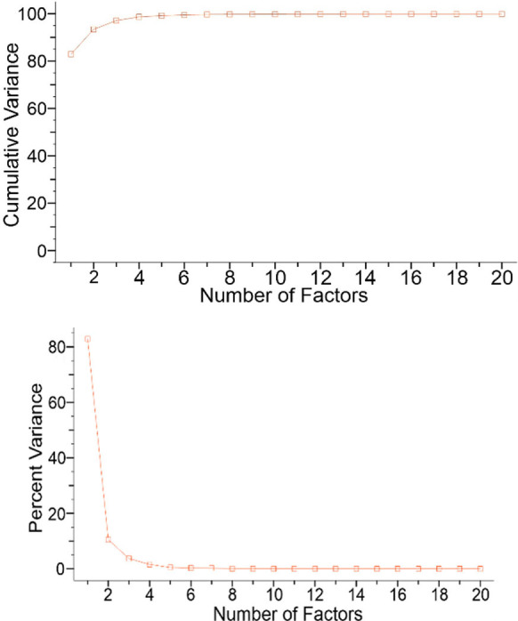
Cumulative variance of Raman dataset (a) and Percent variance value (b).
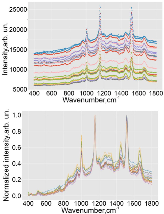
Spectral set for the one probe of healthy volunteer before preprocessing (a) and after it (b).
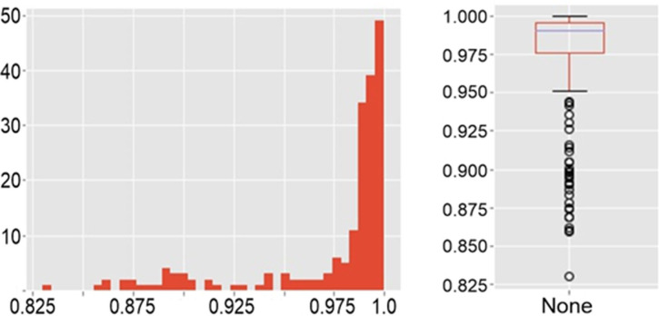
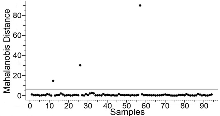
A histogram (a) and a boxplot (b).
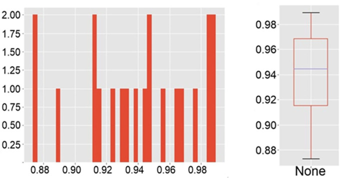
A histogram (a) and a boxplot (b).
References
-
- Roth G. A., Mensah G. A., Johnson C. O., Addolorato G., Ammirati E., Baddour L.M., et al N. C., “Global burden of cardiovascular diseases and risk factors, 1990–2019: update from the GBD 2019 study,” J. Am. Coll. Cardiol. 76(25), 2982–3021 (2020). doi: 10.1016/j.jacc.2020.11.010 - DOI - PMC - PubMed
-
- Furie B., Furie B.C., “Mechanisms of thrombus formation,” N. Engl. J. Med. 359(9), 938–949 (2020). - PubMed
-
- Hartmann J., Hussein A., Trowitzsch E., Becker J., Hennecke K. H., “Treatment of neonatal thrombus formation with recombinant tissue plasminogen activator: six years experience and review of the literature,” Arch. Dis. Child. Fetal Neonatal Ed. 85(1), F18–F22 (2001). doi: 10.1136/fn.85.1.f18 - DOI - PMC - PubMed
-
- Yamada K., Tsuji H., Kimura S., Kato S., Yano S., Ukimura N., et al., “Effects of argatroban and heparin on thrombus formation and tissue plasminogen activator-induced thrombolysis in a microvascular thrombosis model,” Thromb. Res. 109(1), 55–64 (2003). doi: 10.1016/s0049-3848(03)00105-1 - DOI - PubMed
Publication types
MeSH terms
Associated data
LinkOut - more resources
Full Text Sources
Miscellaneous
