

Scleral communication between Glaucoma drainage device capsule and the suprachoroidal space simulating amelanotic choroidal melanoma
- PMID: 35544943
- PMCID: PMC11826707
- DOI: 10.5935/0004-2749.20230073
Scleral communication between Glaucoma drainage device capsule and the suprachoroidal space simulating amelanotic choroidal melanoma
Abstract
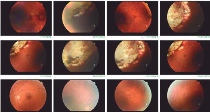
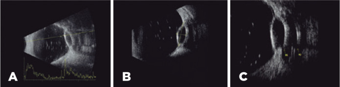
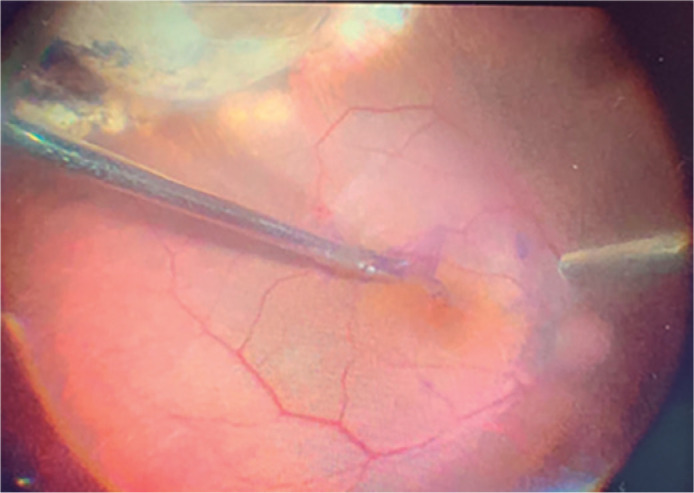
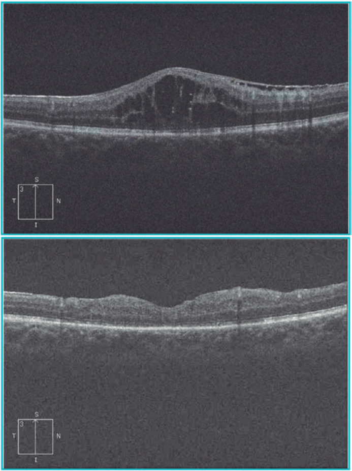
This is a case report involving a 56-year-old male patient with a history of pars plana vitrectomy due to a rhegmatogenous retinal detachment in the right eye that resulted in the implantation of a drainage device after the patient developed secondary glaucoma. Two years after the device's implantation, the patient was referred to our care as his visual acuity had decreased to 20/200 (1.00 LogMAR). At the fundus evaluation, a choroidal amelanotic elevation was observed at the upper temporal equator, and a potential diagnosis was made of amelanotic choroidal melanoma. The ultrasound exam visualized the patient's implanted superotemporal justabulbar drainage device, which revealed a transscleral communication from the plate fibrocapsular's draining space to the suprachoroidal space (fistula). The ultrasound also revealed a focal pocket of choroidal detachment in the patient's superotemporal region, simulating an amelanotic choroidal melanoma. A new pars plana vitrectomy was performed to remove the internal limiting membrane without repercussions at the fistula site. The patient's recovery progressed well, and he regained a visual acuity of 20/70 (0.55 LogMAR). To the best of our knowledge, this is the first case report of this condition.
Relato de caso de paciente 56 anos, sexo masculino, com histórico de vitrectomia via pars plana por descolamento de retina em olho direito e posterior implante de dispositivo de drenagem por glaucoma secundário. Dois anos após o procedimento foi encaminhado ao serviço por baixa de acuidade visual (AV) de 20/200 (1.00 LogMAR). À fundoscopia, observou-se uma elevação amelanótica temporal no equador com hipótese diagnóstica de melanoma de coroide amelanótico. O exame de ultrassom mostrou implante de dispositivo de drenagem justabulbar temporal superior com comunicação transescleral para espaço subcoroidal (fístula), sugerindo bolsão focal de descolamento de coroide em equador temporal superior simulando melanoma de coroide amelanótico. O paciente foi abordado cirurgicamente devido membrana epirretiniana com nova vitrectomia via pars plana para peeling de membrana limitante interna, sem repercussões no local da fístula, evoluindo bem com acuidade visual de 20/70 (0.55 LogMAR). Ao nosso conhecimento, este é o primeiro caso relatado nessa condição.
Conflict of interest statement
Figures
Similar articles
-
Surgical management of silicone oil migrated into suprachoroidal space after vitrectomy.Int J Ophthalmol. 2011;4(4):458-60. doi: 10.3980/j.issn.2222-3959.2011.04.28. Epub 2011 Aug 18. Int J Ophthalmol. 2011. PMID: 22553702 Free PMC article.
-
Suprachoroidal silicone oil migration following 27 gauge 3 ports pars plana vitrectomy - a rare complication and its management.J Pak Med Assoc. 2023 Jun;73(6):1314-1316. doi: 10.47391/JPMA.6823. J Pak Med Assoc. 2023. PMID: 37427640
-
Pars plana vitrectomy in the treatment of combined rhegmatogenous retinal detachment and choroidal detachment in aphakic or pseudophakic patients.Ophthalmic Surg Lasers. 1997 Apr;28(4):288-93. Ophthalmic Surg Lasers. 1997. PMID: 9101566
-
Suprachoroidal hemorrhage during pars plana vitrectomy.Curr Opin Ophthalmol. 2001 Jun;12(3):179-85. doi: 10.1097/00055735-200106000-00006. Curr Opin Ophthalmol. 2001. PMID: 11389343 Review.
-
Pars plana vitrectomy versus scleral buckling for repairing simple rhegmatogenous retinal detachments.Cochrane Database Syst Rev. 2019 Mar 8;3(3):CD009562. doi: 10.1002/14651858.CD009562.pub2. Cochrane Database Syst Rev. 2019. PMID: 30848830 Free PMC article.
References
-
- Tham YC, Li X, Wong TY, Quigley HA, Aung T, Cheng CY. Global prevalence of glaucoma and projections of glaucoma burden through 2040: a systematic review and meta-analysis. Ophthalmology. 2014;121(11):2081–90. Comment in: Ophthalmology. 2015; 122(7): e40-1. Ophthalmology. 2014; 122(7): e41-2. - PubMed
-
- Gedde SJ, Schiffman JC, Feuer WJ, Herndon LW, Brandt JD, Budenz DL, Tube versus Trabeculectomy Study Group Treatment outcomes in the Tube Versus Trabeculectomy (TVT) study after five years of follow-up. Am J Ophthalmol. 2012;153(5):789.e2–803.e2. Comment in: Am J Ophthalmol. 2012; 153(5): 787-8. - PMC - PubMed
-
- Giovingo M. Complications of glaucoma drainage device surgery: a review. Semin Ophthalmol. 2014;29(5-6):397–402. - PubMed
-
- Shin DY, Jung KI, Park HYL, Park CK. Risk factors for choroidal detachment after Ahmed valve implantation in glaucoma patients. Am J Ophthalmol. 2020;211:105–113. - PubMed
Publication types
LinkOut - more resources
Full Text Sources
Miscellaneous
