

SSA-Net: Spatial self-attention network for COVID-19 pneumonia infection segmentation with semi-supervised few-shot learning
- PMID: 35544999
- PMCID: PMC9027296
- DOI: 10.1016/j.media.2022.102459
SSA-Net: Spatial self-attention network for COVID-19 pneumonia infection segmentation with semi-supervised few-shot learning
Abstract
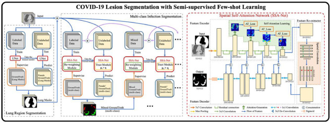
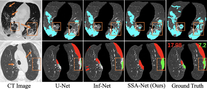
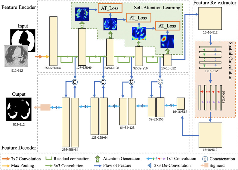
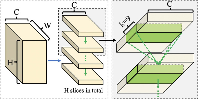
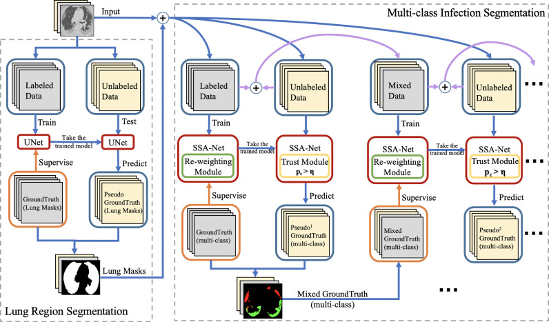
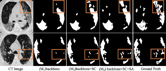
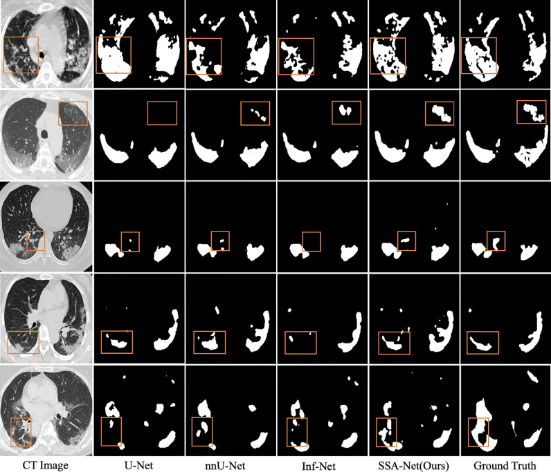
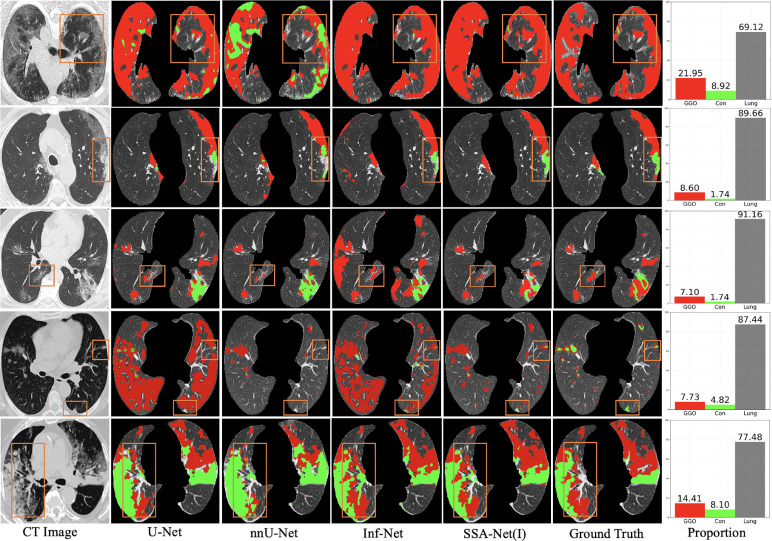
Coronavirus disease (COVID-19) broke out at the end of 2019, and has resulted in an ongoing global pandemic. Segmentation of pneumonia infections from chest computed tomography (CT) scans of COVID-19 patients is significant for accurate diagnosis and quantitative analysis. Deep learning-based methods can be developed for automatic segmentation and offer a great potential to strengthen timely quarantine and medical treatment. Unfortunately, due to the urgent nature of the COVID-19 pandemic, a systematic collection of CT data sets for deep neural network training is quite difficult, especially high-quality annotations of multi-category infections are limited. In addition, it is still a challenge to segment the infected areas from CT slices because of the irregular shapes and fuzzy boundaries. To solve these issues, we propose a novel COVID-19 pneumonia lesion segmentation network, called Spatial Self-Attention network (SSA-Net), to identify infected regions from chest CT images automatically. In our SSA-Net, a self-attention mechanism is utilized to expand the receptive field and enhance the representation learning by distilling useful contextual information from deeper layers without extra training time, and spatial convolution is introduced to strengthen the network and accelerate the training convergence. Furthermore, to alleviate the insufficiency of labeled multi-class data and the long-tailed distribution of training data, we present a semi-supervised few-shot iterative segmentation framework based on re-weighting the loss and selecting prediction values with high confidence, which can accurately classify different kinds of infections with a small number of labeled image data. Experimental results show that SSA-Net outperforms state-of-the-art medical image segmentation networks and provides clinically interpretable saliency maps, which are useful for COVID-19 diagnosis and patient triage. Meanwhile, our semi-supervised iterative segmentation model can improve the learning ability in small and unbalanced training set and can achieve higher performance.
Keywords: COVID-19; Few-shot learning; Lesion segmentation; Semi-supervised.
Copyright © 2022. Published by Elsevier B.V.
Conflict of interest statement
Declaration of Competing Interest The authors declare that they have no known competing financial interests or personal relationships that could have appeared to influence the work reported in this paper.
Figures
Similar articles
-
Inf-Net: Automatic COVID-19 Lung Infection Segmentation From CT Images.IEEE Trans Med Imaging. 2020 Aug;39(8):2626-2637. doi: 10.1109/TMI.2020.2996645. IEEE Trans Med Imaging. 2020. PMID: 32730213
-
CARes-UNet: Content-aware residual UNet for lesion segmentation of COVID-19 from chest CT images.Med Phys. 2021 Nov;48(11):7127-7140. doi: 10.1002/mp.15231. Epub 2021 Sep 25. Med Phys. 2021. PMID: 34528263 Free PMC article.
-
A modality-collaborative convolution and transformer hybrid network for unpaired multi-modal medical image segmentation with limited annotations.Med Phys. 2023 Sep;50(9):5460-5478. doi: 10.1002/mp.16338. Epub 2023 Mar 15. Med Phys. 2023. PMID: 36864700
-
Learning with limited annotations: A survey on deep semi-supervised learning for medical image segmentation.Comput Biol Med. 2024 Feb;169:107840. doi: 10.1016/j.compbiomed.2023.107840. Epub 2023 Dec 16. Comput Biol Med. 2024. PMID: 38157773 Review.
-
A review of self-supervised, generative, and few-shot deep learning methods for data-limited magnetic resonance imaging segmentation.NMR Biomed. 2024 Aug;37(8):e5143. doi: 10.1002/nbm.5143. Epub 2024 Mar 24. NMR Biomed. 2024. PMID: 38523402 Review.
Cited by
-
Longitudinally consistent registration and parcellation of cortical surfaces using semi-supervised learning.Med Image Anal. 2024 Aug;96:103193. doi: 10.1016/j.media.2024.103193. Epub 2024 May 7. Med Image Anal. 2024. PMID: 38823362 Free PMC article.
-
Lung and Infection CT-Scan-Based Segmentation with 3D UNet Architecture and Its Modification.Healthcare (Basel). 2023 Jan 10;11(2):213. doi: 10.3390/healthcare11020213. Healthcare (Basel). 2023. PMID: 36673581 Free PMC article.
-
Few-shot segmentation with duplex network and attention augmented module.Front Neurorobot. 2023 Jun 21;17:1206189. doi: 10.3389/fnbot.2023.1206189. eCollection 2023. Front Neurorobot. 2023. PMID: 37416851 Free PMC article.
-
Semi-supervised Segmentation of Histopathology Images with Noise-Aware Topological Consistency.Comput Vis ECCV. 2024;15136:271-289. doi: 10.1007/978-3-031-73229-4_16. Epub 2024 Oct 25. Comput Vis ECCV. 2024. PMID: 40557360 Free PMC article.
-
Dual-branch Transformer for semi-supervised medical image segmentation.J Appl Clin Med Phys. 2024 Oct;25(10):e14483. doi: 10.1002/acm2.14483. Epub 2024 Aug 12. J Appl Clin Med Phys. 2024. PMID: 39133901 Free PMC article.
References
-
- Apostolopoulos S., De Zanet S., Ciller C., Wolf S., Sznitman R. International Conference on Medical Image Computing and Computer-Assisted Intervention. Springer; 2017. Pathological OCT retinal layer segmentation using branch residual U-shape networks; pp. 294–301.
-
- Chen N., Zhou M., Dong X., Qu J., Gong F., Han Y., Qiu Y., Wang J., Liu Y., Wei Y., Xia J., Yu T., Zhang X., Zhang L. Epidemiological and clinical characteristics of 99 cases of 2019 novel coronavirus pneumonia in Wuhan, China: a descriptive study. Lancet. 2020;395(10223):507–513. doi: 10.1016/S0140-6736(20)30211-7. - DOI - PMC - PubMed
Publication types
MeSH terms
LinkOut - more resources
Full Text Sources
Medical
Research Materials
