. 2022;14(6):643-656.
doi: 10.1159/000524560. Epub 2022 May 11.

IL-1 Mediates Tissue-Specific Inflammation and Severe Respiratory Failure in COVID-19

Affiliations

IL-1 Mediates Tissue-Specific Inflammation and Severe Respiratory Failure in COVID-19

Georgios Renieris et al. J Innate Immun. 2022.

Abstract

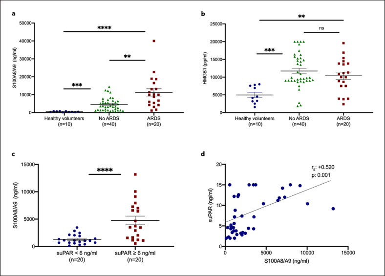
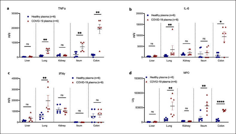
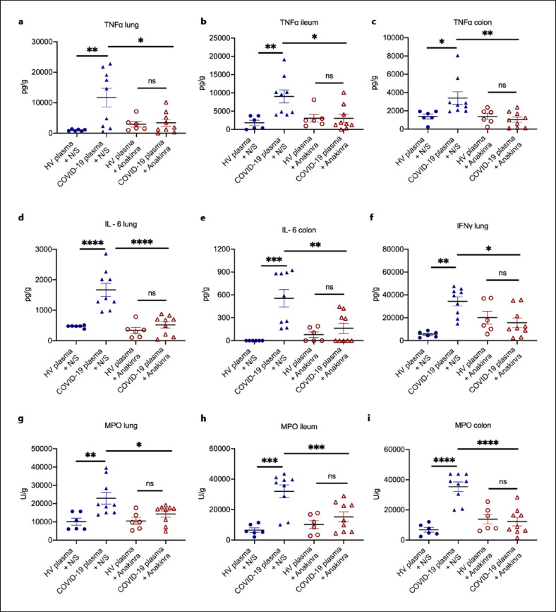
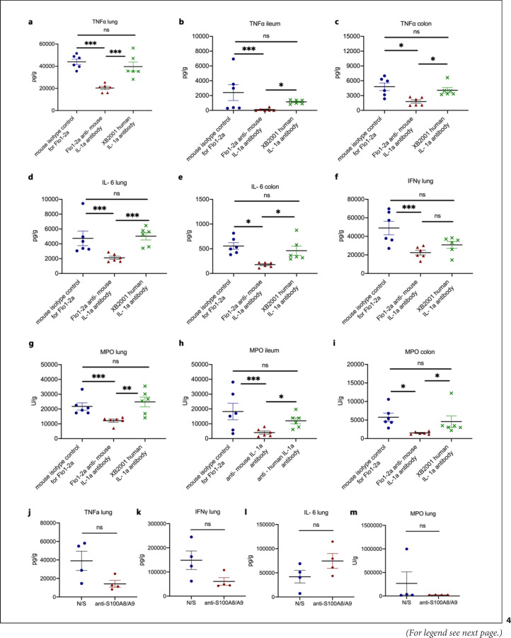
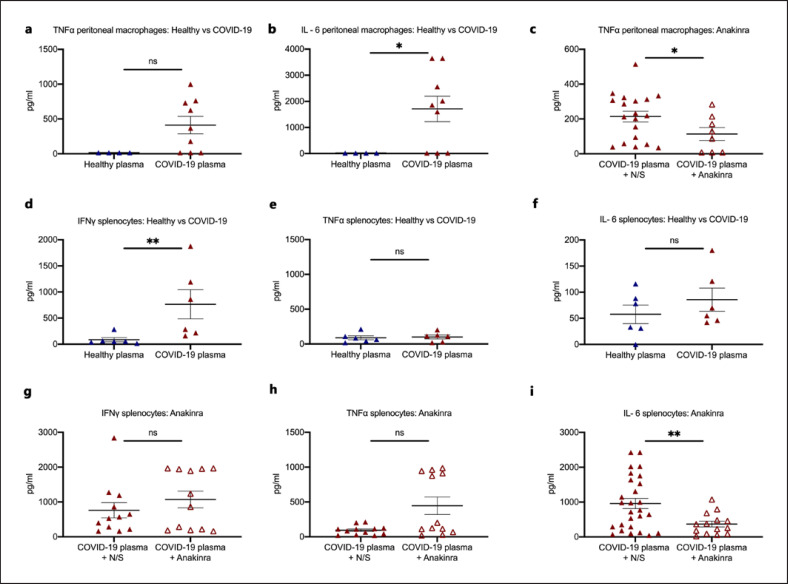
Acute respiratory distress syndrome (ARDS) in COVID-19 has been associated with catastrophic inflammation. We present measurements in humans and a new animal model implicating a role in danger-associated molecular patterns. Calprotectin (S100A8/A9) and high-mobility group box 1 (HMGB1) were measured in patients without/with ARDS, and admission calprotectin was associated with soluble urokinase plasminogen activator receptor (suPAR). An animal model was developed by intravenous injection of plasma from healthy or patients with COVID-19 ARDS into C57/BL6 mice once daily for 3 consecutive days. Mice were treated with one anti-S100A8/A9 antibody, the IL-1 receptor antagonist anakinra or vehicle, and Flo1-2a anti-murine anti-IL-1α monoclonal antibody or the specific antihuman IL-1α antibody XB2001 or isotype controls. Cytokines and myeloperoxidase (MPO) were measured in tissues. Calprotectin, but not HMGB1, was elevated in ARDS. Higher suPAR indicated higher calprotectin. Animal challenge with COVID-19 plasma led to inflammatory reactions in murine lung and intestines as evidenced by increased levels of TNFα, IL-6, IFNγ, and MPO. Lung inflammation was attenuated with anti-S100A8/A9 pre-treatment. Anakinra treatment restored these levels. Similar decrease was found in mice treated with Flo1-2a but not with XB2001. Circulating alarmins, specifically calprotectin, of critically ill COVID-19 patients induces tissue-specific inflammatory responses through an IL-1-mediated mechanism. This could be attenuated through inhibition of IL-1 receptor or of IL-1α.

Trial registration: ClinicalTrials.gov NCT04357366.

Keywords: Acute respiratory distress syndrome; Calprotectin; Coronavirus diseases 2019; Interleukin-1; Interleukin-6; Severe respiratory failure; Soluble urokinase plasminogen activator receptor; Tumor necrosis factor.


Conflict of interest statement

J. Eugen-Olsen is a cofounder, shareholder, and CSO of ViroGates A7S, Denmark, and is named inventor on patents on suPAR owned by Copenhagen University Hospital Hvidovre, Denmark. He is granted by the Horizon 2020 European Grant RISCinCOVID. J. Simard is the CEO and founder of XBiotech. M.G. Netea is a scientific founder of TTxD and received research grants from GSK and ViiV Healthcare. E.J. Giamarellos-Bourboulis has received honoraria from AbbVie USA, Abbott CH, InflaRx GmbH, MSD Greece, XBiotech Inc., and Angelini Italy; independent educational grants from AbbVie, Abbott, Astellas Pharma Europe, Axis Shield, bioMérieux Inc, InflaRx GmbH, Sobi, and XBiotech Inc.; and funding from the FrameWork 7 program HemoSpec (granted to the National and Kapodistrian University of Athens), the Horizon 2020 Marie-Curie Project European Sepsis Academy (granted to the National and Kapodistrian University of Athens), and the Horizon 2020 European Grant ImmunoSep (granted to the Hellenic Institute for the Study of Sepsis).

Figures

Fig. 1
Fig. 1
Calprotectin is increased in patients with ARDS due to COVID-19. a, b Concentrations of calprotectin (S100A8/A9) and HMGB1 were measured in the plasma from HVs, from patients with COVID-19 who did not develop ARDS during follow-up on the day of hospital admission, and from patients with ARDS by COVID-19 on the first day of ARDS. Comparison by the Mann-Whitney U test; * p < 0.05; ** p < 0.01; *** p < 0.001; ****; p < 0.0001. c, d suPAR was measured in 40 patients screened for participation in the SAVE trial (3). None of the patients with suPAR less than 6 ng/mL developed ARDS, whereas 6 patients (30%) with suPAR 6 ng/mL or more developed ARDS. c Concentrations of calprotectin in 20 patients with suPAR less than 6 ng/mL and in 20 patients with suPAR 6 ng/mL or more. Comparison by the Mann-Whitney U test; * p < 0.05. d Correlation of levels of suPAR with plasma calprotectin on day of admission Spearman rank correlation coefficients (rs), interpolation lines, and p values are provided. ns, nonsignificant.
Fig. 2
Fig. 2
A COVID-19-like animal model of compartmentalized hyperinflammation. C57Bl6 mice were injected i.v. with plasma of HVs or patients with ARDS due to COVID-19 for 3 consecutive days. Mice were sacrificed on day 4. Tissue concentrations of TNFα (a), IL-6 (b), IFNγ (c), and MPO activity (d) were determined. Comparison by the Mann-Whitney U test; * p < 0.05; ** p < 0.01; **** p < 0.0001. ns, nonsignificant.
Fig. 3
Fig. 3
IL-1 inhibition attenuates the compartmentalized hyperinflammation in a COVID-19-like murine model. In a COVID-19-like infection model, C57Bl6 mice were challenged i.v. with plasma of HVs or patients with ARDS due to COVID-19 for 3 consecutive days. In separate experiments, on each day of plasma challenge, mice were treated with anakinra, which inhibits human and murine IL-1, or vehicle. Mice were sacrificed on day 4. a–c TNFα, IL-6 (d, e), IFNγ (f), and MPO activity (g–i) was determined in tissues. Comparison by the Mann-Whitney U test; * p < 0.05; ** p < 0.01. ns nonsignificant.
Fig. 4
Fig. 4
Murine IL-1α and S100A8/A9 drive the hyperinflammation caused by SARS-CoV-2. In a COVID-19-like infection model, C57Bl6 mice were challenged i.v. with plasma of patients with ARDS due to COVID-19 for 3 consecutive days. In separate experiments, on each day of plasma challenge, mice were treated with Flo1-2a anti-murine IL-1α antibody or XB2001 human IL-1α antibody or murine isotype control for Flo1-2a or pre-treated with anti-S100A8/A9. Mice were sacrificed on day 4. a–c TNFα in the lung, ileum, and colon and modulation by blockade of human or murine IL-1α. d, e IL-6 in the lung, ileum, and colon and modulation by blockade of human or murine IL-1α. f IFNγ in the lung, ileum, and colon and modulation by blockade of human or murine IL-1α. g–i MPO activity in the lung, ileum, and colon and modulation by blockade of human or murine IL-1α. j TNFα in the lung and modulation by blockade of S100A8/A9. k IFNγ in the lung and modulation by blockade of S100A8/A9. l IL-6 in the lung and modulation by blockade of S100A8/A9. m MPO activity in the lung and modulation by blockade of S100A8/A9. Comparison by the Mann-Whitney U test; * p < 0.05; ** p < 0.01; *** p < 0.001. ns, nonsignificant.
Fig. 5
Fig. 5
Activation of peritoneal macrophages and splenocytes and modulation through IL-1 inhibition. a, b In a COVID-19-like infection model, C57Bl6 mice were treated i.v. with plasma of HVs or patients with ARDS due to COVID-19 for 3 consecutive days. Mice were sacrificed on day 4. Peritoneal macrophages were isolated and incubated with bacterial LPS for the production of TNFα and IL-6. c In a COVID-19-like infection model, C57Bl6 mice were treated i.v. with plasma of patients with ARDS due to COVID-19 with or without treatment with the IL-1 receptor inhibitor anakinra for 3 consecutive days. Mice were sacrificed on day 4. Peritoneal macrophages were isolated and incubated with bacterial LPS for the production of TNFα. d–f In a COVID-19-like infection model, C57Bl6 mice were treated i.v. with plasma of HVs or patients with ARDS due to COVID-19 for 3 consecutive days. Mice were sacrificed on day 4. Splenocytes were isolated and incubated with heat-killed C. albicans for the production of IFNγ, TNFα, and IL-6. e–i In a COVID-19-like infection model, C57Bl6 mice were treated i.v. with plasma of patients with ARDS due to COVID-19 with or without treatment with the IL-1 receptor inhibitor anakinra for 3 consecutive days. Mice were sacrificed on day 4. Splenocytes were isolated and incubated with heat-killed C. albicans for the production of IFNγ, TNFα, and IL-6. Comparison by the Mann-Whitney U test; * p < 0.05; ** p < 0.01. LPS, lipopolysaccharide; ns nonsignificant.
Fig. 6
Fig. 6
COVID-19-like hyperinflammation is associated with susceptibility to A. baumannii infections. a C57Bl6 mice were injected i.v. with plasma of HVs or patients with ARDS due to COVID-19 for 3 consecutive days. On day 4, mice were challenged i.p. with E. coli or A. baumannii. Survival comparison by the log-rank test and the respective p value is provided. b, c C57Bl6 mice were injected i.v. with plasma of patients with ARDS due to COVID-19 for 3 consecutive days. On each day of plasma challenge, half of the mice were treated with anakinra. On day 4, mice were challenged i.p. with E. coli (b) or A. baumannii (c). Survival comparison by the log-rank test and the respective p value is provided.

References

    1. Giamarellos-Bourboulis EJ, Netea MG, Rovina N, Akinosoglou K, Antoniadou A, Antonakos N, et al. Complex immune dysregulation in COVID-19 patients with severe respiratory failure. Cell Host Microbe. 2020;27:992–1000.e3. - PMC - PubMed
    1. Rodrigues TS, de Sá KSG, Ishimoto AY, Becerra A, Oliveira S, Almeida L, et al. Inflammasomes are activated in response to SARS-CoV-2 infection and are associated with COVID-19 severity in patients. J Exp Med. 2021;218:e20201707. - PMC - PubMed
    1. Kyriazopoulou E, Panagopoulos P, Metallidis S, Dalekos GN, Poulakou G, Gatselis N, et al. An open label trial of anakinra to prevent respiratory failure in COVID-19. eLife. 2021;10:e66125. - PMC - PubMed
    1. de Luca A, Smeekens SP, Casagrande A, Iannitti R, Conway KL, Gresnigt MS, et al. IL-1 receptor blockade restores autophagy and reduces inflammation in chronic granulomatous disease in mice and in humans. Proc Natl Acad Sci U S A. 2014;111:3526–31. - PMC - PubMed
    1. Aeffner F, Bolon B, Davis IC. Mouse models of acute respiratory distress syndrome: a review of analytical approaches, pathologic features, and common measurements. Toxicol Pathol. 2015;43:1074–92. - PubMed

Publication types

Substances

Associated data