. 2022 May 10;39(6):110791.
doi: 10.1016/j.celrep.2022.110791.

Functional genome-wide short hairpin RNA library screening identifies key molecules for extracellular vesicle secretion from microglia

Affiliations

Functional genome-wide short hairpin RNA library screening identifies key molecules for extracellular vesicle secretion from microglia

Zhi Ruan et al. Cell Rep. .

Abstract

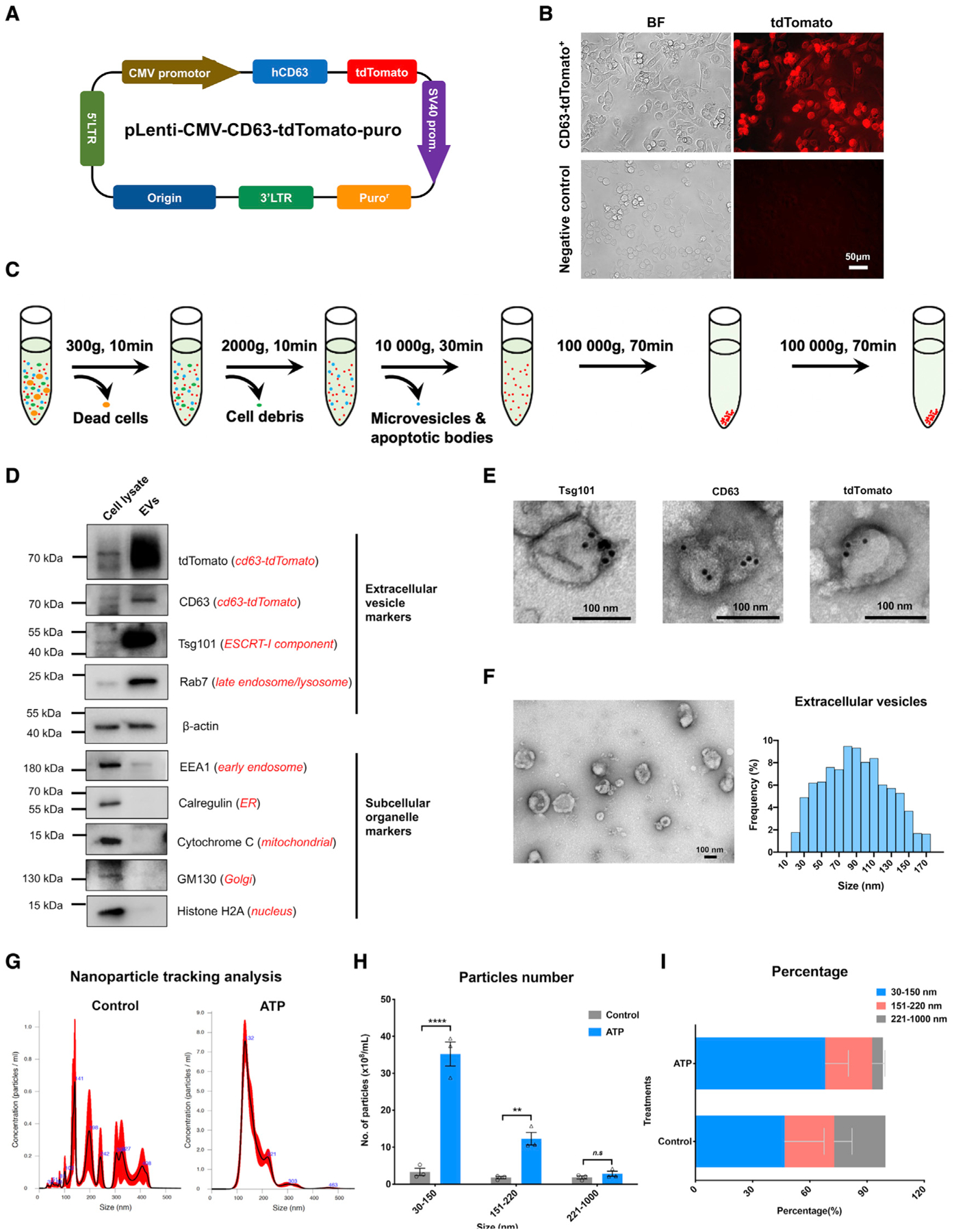
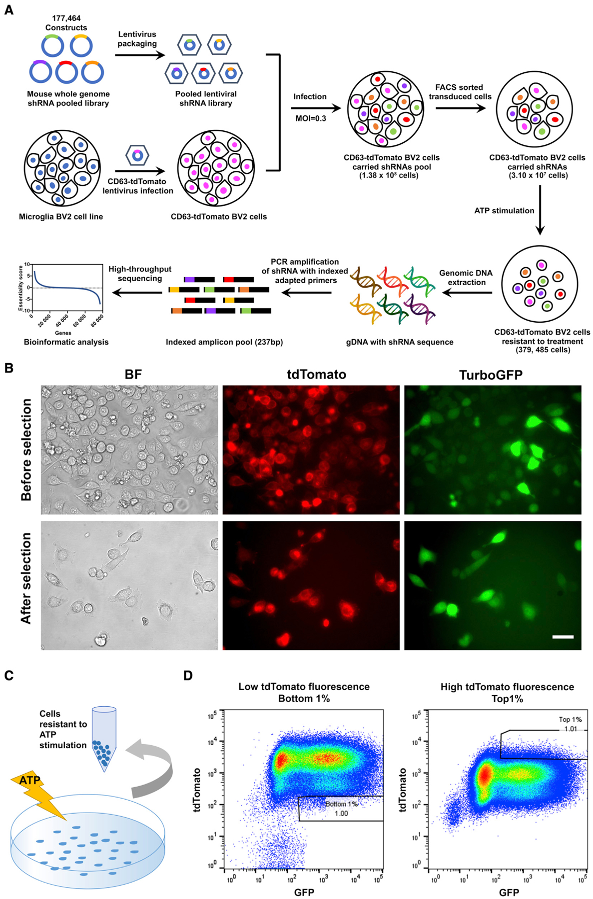
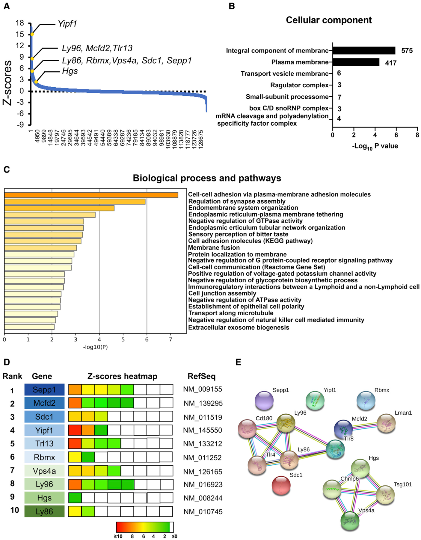
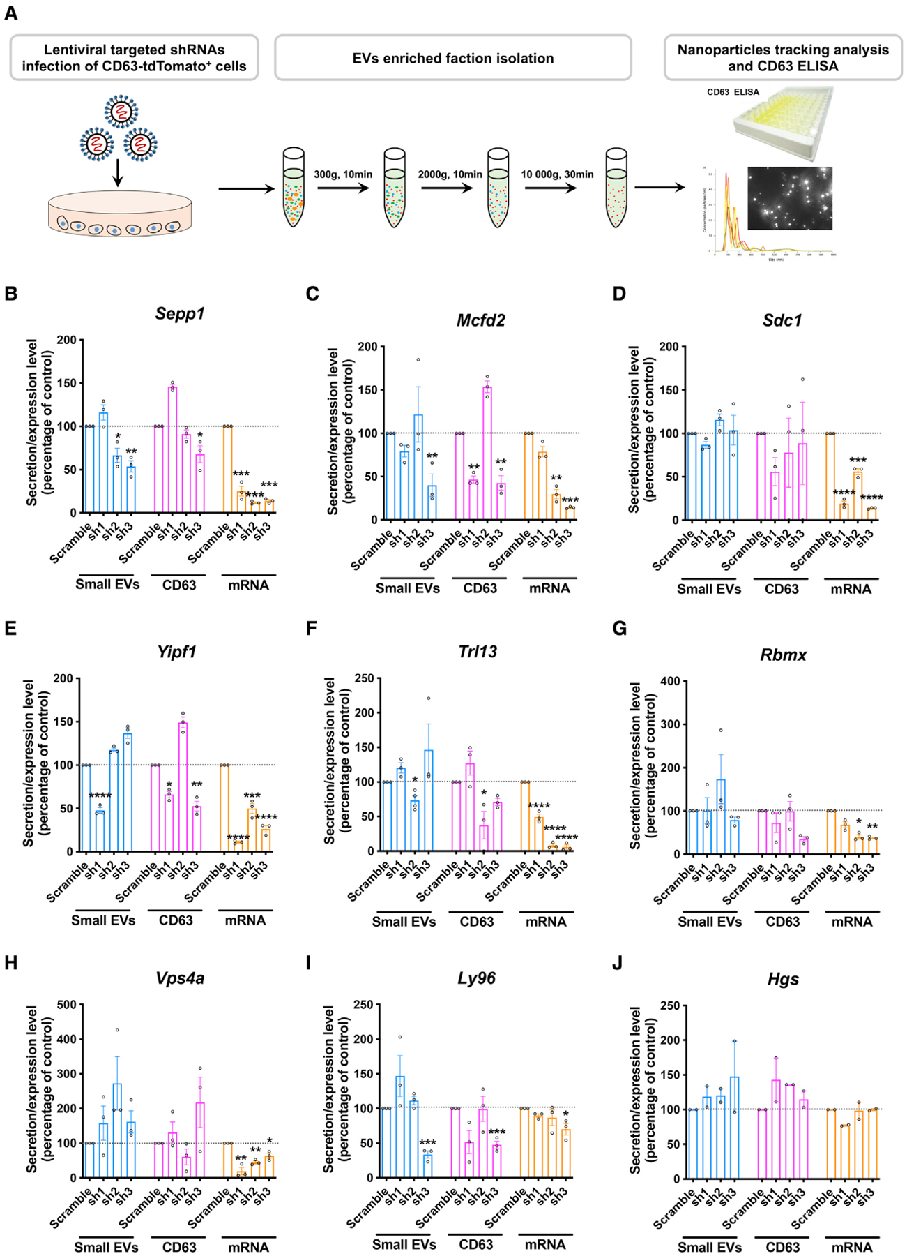
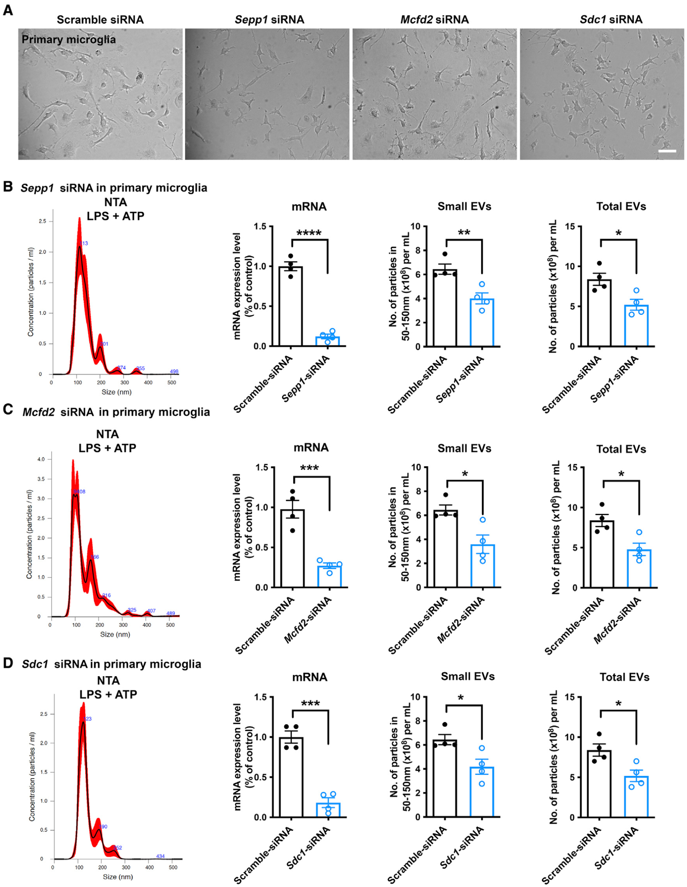
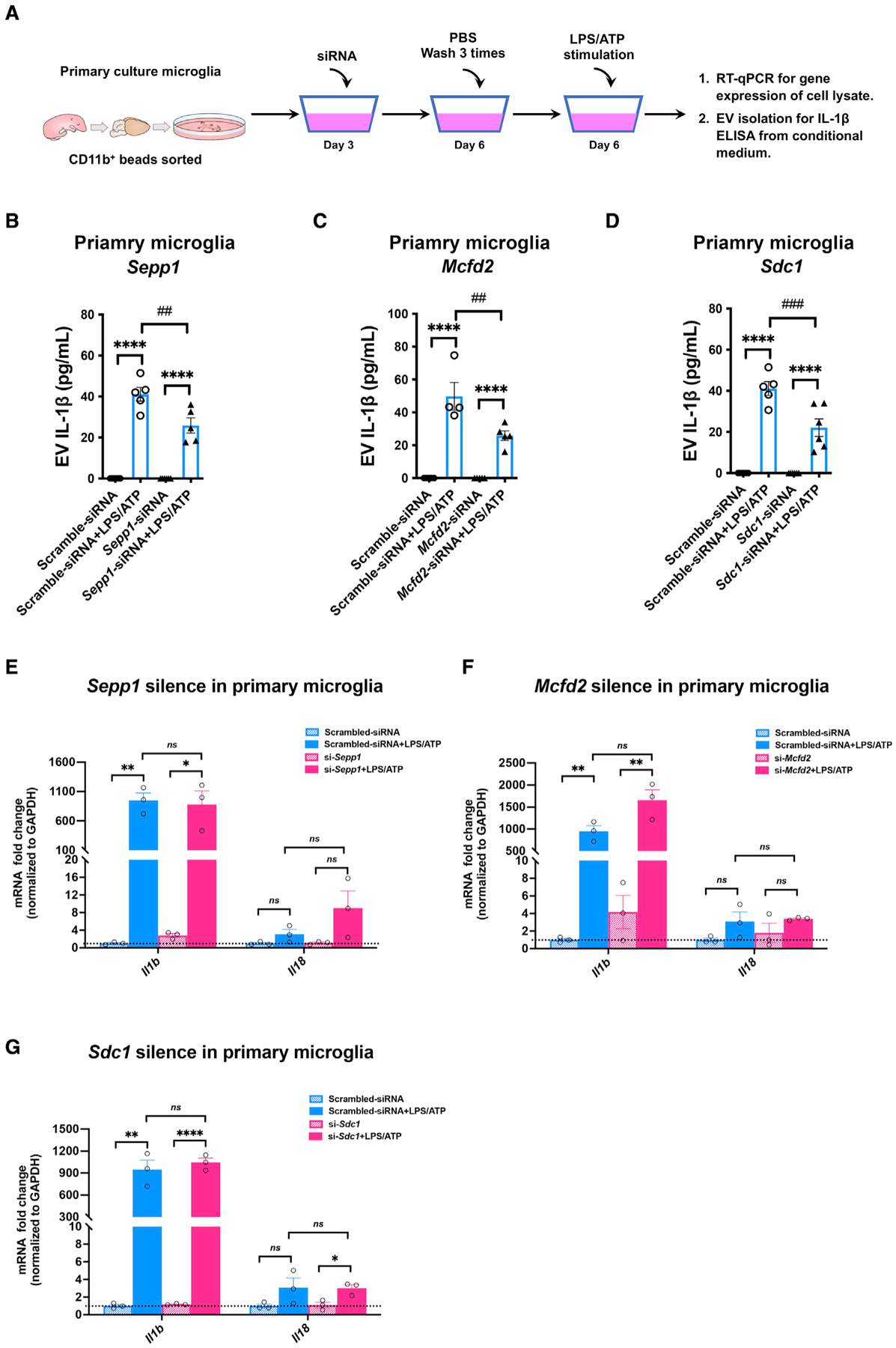
Activated microglia release extracellular vesicles (EVs) as modulators of brain homeostasis and innate immunity. However, the molecules critical for regulating EV production from microglia are poorly understood. Here we establish a murine microglial cell model to monitor EV secretion by measuring the fluorescence signal of tdTomato, which is linked to tetraspanin CD63. Stimulation of tdTomato+ cells with ATP induces rapid secretion of EVs and a reduction in cellular tdTomato intensity, reflecting EV secretion. We generate a GFP+ tdTomato+ cell library expressing TurboGFP and barcoded short hairpin RNAs for genome-wide screening using next-generation sequencing. We identify Mcfd2, Sepp1, and Sdc1 as critical regulators of ATP-induced EV secretion from murine microglia. Small interfering RNA (siRNA-based) silencing of each of these genes suppresses lipopolysaccharide- and ATP-induced inflammasome activation, as determined by interleukin-1β release from primary cultured murine microglia. These molecules are critical for microglial EV secretion and are potential therapeutic targets for neuroinflammatory disorders.

Keywords: CP: Neuroscience; extracellular vesicle; interleukin-1β; microglia; microtubule-associated protein tau; neuroinflammation; shRNA library screening.


Conflict of interest statement

Declarations of interests T.I. had a sponsored research agreement with Ono Pharmaceutical and consults with Takeda.

Figures

Figure 1.
Figure 1.. Creation of the tdTomato-CD63+ BV-2 stable cell line and characterization of EV release after ATP stimulation
(A) Arrangement of the engineered pLenti-tdTomato-CD63 vector. (B) Creation of the tdTomato-CD63+ reporter BV-2 stable cell line. Scale bar, 50 μm. (C) Workflow to purify the small EV fraction from the conditioned culture medium of the tdTomato-CD63+ BV-2 stable cell line. (D) Representative immunoblots of typical protein markers for small EVs, engineered small EV tags, and other subcellular organelles from cell lysate and purified small EV fractions of conditioned medium. (E) Representative immunogold electron microscopy images of Tsg101, CD63, and tdTomato labeling on small EVs isolated from conditioned medium. Scale bar, 100 nm. (F) Representative transmission electron microscopy images (left panel) and size distribution (right panel) of exosomes isolated from conditioned medium. In total, 16 images were analyzed. Scale bar, 100 nm. (G and H) Representative size distribution of small EVs isolated from conditioned medium of tdTomato-CD63+ BV-2 cells with or without ATP stimulation by Nanosight NTA. Two-tailed Student’s t test; **p < 0.01; ****p < 0.0001; ns, no statistical significance; compared with the scramble-shRNA group; n = 3 independent experiments, each sample was recorded for 4 videos. Graphs indicate mean ± SEM. (I) The percentage distribution of different size fractions of EVs isolated from the conditioned medium.
Figure 2.
Figure 2.. Positive cells containing silencing factors acting during EV secretion
(A) Schematic of the workflow of genome-wide shRNA library screening. A genome-wide mouse shRNA library containing 177,464 constructs was packed into lentiviral particles and transduced into tdTomato-CD63 overexpressing BV-2 cells at a low multiplicity of infection (MOI). The shRNA-transduced cells were sorted by FACS to generate a mutant cell pool. Mutant cells were amplified and treated with ATP stimulation for genetic screening. Genomic DNA was extracted from the treated cells, and the shRNA fragment was amplified by PCR. The copy number of shRNAs was determined by HTS and bioinformatics analysis. (B) Transduced tdTomato-CD63 and shRNAs lentiviral BV-2 cells sorted by FACS. (C) The workflow of ATP stimulation and cell collection. (D) FACS gating panels of the bottom 1% of cells representing cells sensitive to ATP stimulation for EV secretion (left) and top 1% of cells representing cells resistant to ATP stimulation for EV secretion. In total, 18 cell pools were collected.
Figure 3.
Figure 3.. Identification of top-ranked candidates from the shRNA library screening
(A) Negative control normalized Z scores of all pooled RNAi screening data, ordered from most negative to positive. (B) The seven most significantly enriched GO terms from the list of screened hits (p < 0.05) after Benjamini-Hochberg multiple-testing correction. (C) Biological processes and pathways identified by Metascape with the candidate genes from the integral component of the membrane and plasma membrane. (D) The 10 top-ranked candidates from the shRNA screen. The enrichment in cells sorted by FACS (Z score) of the corresponding hairpins is shown using a colored heatmap, where each square represents an independent hairpin. Crosses indicate excluded hairpins for which the read number was below a set threshold. The candidates were ranked based on the Z score for the hairpins targeting the same transcript. Factors linked to the same complexes/pathways are color coded. (E) Candidate genes with GO terms including “secretion” and interactions by STRING association.
Figure 4.
Figure 4.. Modulation of EV secretion by silencing selected gene targets
(A) Workflow to validate a potential “hit” by NTA and CD63 ELISA. (B–J) EV and CD63 secretion and specific mRNA levels in cells transduced with lentiviral shRNA clones targeting Sepp1 (B), Mcfd2 (C), Sdc1 (D), Yipf1 (E), Tlr13 (F), Rbmx (G), Vps4a (H), Ly96 (I), and Hgs (J). *p < 0.05, **p < 0.01, ***p < 0.001, ****p < 0.0001 compared with the scramble shRNA group, as determined by one-way ANOVA (alpha = 0.05) and Tukey’s post hoc test. n = 3 independent experiments, a technical duplicate analysis for each sample in CD63 ELISA and qRT-PCR. Graphs indicate mean ± SEM.
Figure 5.
Figure 5.. Validation of EVs secreted by Mcfd2, Sepp1, and Sdc1 knockdown primary cultured murine microglia
(A) Primary microglia transfected with siRNA targeting Sepp1, Mcfd2, and Sdc1. (B–D) Particle distribution, specific mRNA levels, and EV and total particle secretion in primary microglia transduced with siRNAs targeting Sepp1 (B), Mcfd2 (C), and Sdc1 (D). Two-tailed Student’s t test, *p < 0.05, **p < 0.01, ***p < 0.001, ****p < 0.0001 compared with the scramble shRNA group; n = 4 independent experiment, each sample recorded with 4 videos. Graphs indicate mean ± SEM.
Figure 6.
Figure 6.. IL-1β secretion in EVs from primary murine microglia
(A) The workflow for measuring pro-inflammatory cytokine IL-1β secretion from microglia after siRNA silencing. (B–D) EV-associated IL-1β from primary microglia transduced with siRNAs targeting Sepp1 (B), Mcfd2 (C), and Sdc1 (D). *p < 0.05, **p < 0.01, ***p < 0.001, ****p < 0.0001 compared with the control group; ##p < 0.01, ###p < 0.001 compared with the scramble group; determined by two-way ANOVA (alpha = 0.05) and Tukey’s post hoc test; n = 4–5 independent experiments. Graphs indicate mean ± SEM. (E–G) mRNA expression of Il1b and Il18 in primary cultured murine microglia after siRNA-based silencing of Sepp1 (E), Sdc1 (F), and Mcfd2 (G); treated with vehicle or LPS/ATP; and tested for mRNA expression of Il1b and Il18. *p < 0.05, **p < 0.01, ***p < 0.001, ****p < 0.0001 compared with the control group; determined by two-way ANOVA (alpha = 0.05) and Tukey’s post hoc test; n = 3 independent experiments. Graphs indicate mean ± SEM.

References

    1. Andrei C, Dazzi C, Lotti L, Torrisi MR, Chimini G, and Rubartelli A (1999). The secretory route of the leaderless protein interleukin 1beta involves exocytosis of endolysosome-related vesicles. Mol. Biol. Cell 10, 1463–1475. 10.1091/mbc.10.5.1463. - DOI - PMC - PubMed
    1. Andreu Z, and Yanez-Mo M (2014). Tetraspanins in extracellular vesicle formation and function. Front Immunol. 5, 442. 10.3389/fimmu.2014.00442. - DOI - PMC - PubMed
    1. Asai H, et al. (2015). Depletion of microglia and inhibition of exosome synthesis halt tau propagation. Nat Neurosci 18, 1584–1593. 10.1038/nn.4132. - DOI - PMC - PubMed
    1. Baietti MF, Zhang Z, Mortier E, Melchior A, Degeest G, Geeraerts A, Ivarsson Y, Depoortere F, Coomans C, Vermeiren E, et al. (2012). Syndecan-syntenin-ALIX regulates the biogenesis of exosomes. Nat. Cell Biol 14, 677–685. 10.1038/ncb2502. - DOI - PubMed
    1. Bartels T, De Schepper S, and Hong S (2020). Microglia modulate neurodegeneration in Alzheimer’s and Parkinson’s diseases. Science 370, 66–69. 10.1126/science.abb8587. - DOI - PubMed

Publication types