. 2022 Jan 28;47(1):79-85.
doi: 10.11817/j.issn.1672-7347.2022.211227.

Outcome comparison of sural neurofasciocutaneous flap for reconstructing soft tissue defects in forefoot and around ankle

[Article in English, Chinese]
Affiliations

Outcome comparison of sural neurofasciocutaneous flap for reconstructing soft tissue defects in forefoot and around ankle

[Article in English, Chinese]
Lihong Liu et al. Zhong Nan Da Xue Xue Bao Yi Xue Ban. .

Abstract

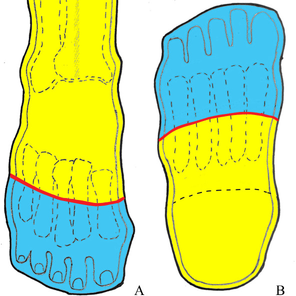
Objectives: To summarize our experience with the sural neurofasciocutaneous flap for reconstructing the soft tissue defects over the forefoot distal to the connecting line of midpoints in the metatarsal bones, and to compare the outcomes between the flap for resurfacing the defects distal and proximal to the connecting line.

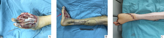
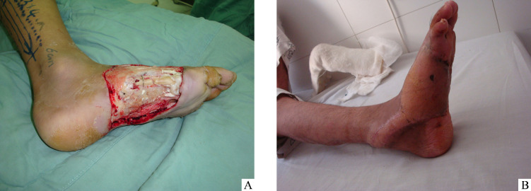
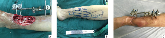
Methods: The clinical data of 425 sural neurofasciocutaneous flaps for repairing the soft tissue defects in the middle and lower leg, ankle, and foot between Apr. 2002 and Apr. 2020 were reviewed. Based on the connecting line of midpoints of the metatarsals, the sural neurofasciocutaneous flaps were divided into a forefoot group (flaps with furthest edges distal to the connecting line) and a peri-ankle group (flaps with the furthest edges proximal to the connecting line).

Results: The partial necrosis rate in the forefoot group (14.5%, 10/69) was significantly higher than that in the peri-ankle group (7.0%, 25/356), with significant difference (P<0.05). Using the flap alone or in combination with a simple salvage treatment, the ratio of successful coverages of the defects was 98.6% (68/69) in the forefoot group, and 97.8% (348/356) in the peri-ankle group, respectively, with no statistical difference (P>0.05).

Conclusions: The sural neurofasciocutaneous flap is a better choice for covering the soft tissue defects over the forefoot distal to the connecting line of midpoints of the metatarsal bones. The survival reliability of the sural neurofasciocutaneous flap reconstructing the soft tissue defect proximal to the connecting line is superior to that of the flap reconstructing the defect distal to the connecting line.

目的: 总结腓肠神经营养血管皮瓣修复跖骨中点连线以远前足创面的临床经验,比较该皮瓣修复跖骨中点连线以远及以近创面的临床疗效。方法: 回顾性分析2002年4月至2020年4月间修复小腿中下段、踝及足部皮肤软组织缺损的425例腓肠神经营养血管皮瓣的临床资料。以跖骨中点连线为界,将腓肠神经营养血管皮瓣分为前足组(皮瓣最远端位于该连线以远的皮瓣)和踝周组(皮瓣最远端位于该连线以近的皮瓣)。结果: 前足组皮瓣部分坏死率(14.5%,10/69)高于踝周组(7.0%,25/356),两组比较差异有统计学意义(P<0.05)。前足组和踝周组单独应用该皮瓣或联合简单二期处理覆盖创面的成功率分别为98.6%(68/69)和97.8%(348/356),两组比较差异无统计学意义(P>0.05)。结论: 腓肠神经营养血管皮瓣是修复跖骨中点连线以远的前足皮肤软组织缺损的一种较理想的方法,该皮瓣修复跖骨中点连线以近创面的成活可靠性优于修复跖骨中点连线以远创面。.

Keywords: perforator flap; peroneal artery; sural nerve; surgical flap.


Conflict of interest statement

作者声称无任何利益冲突。

Figures

图1
图1
前足及踝周划分示意图 Figure 1 Illustration of demarcation of forefoot and peri-ankle A: Anterior view; B: Planta pedis view. With the connecting line (red line) of midpoints of metatarsals as boundary, the middle and lower leg, ankle and foot are divided into two regions: forefoot region (distal to the connecting line, blue area) and peri-ankle region (proximal to the connecting line, yellow area).
图2
图2
53岁男性患者外伤致左足背皮肤软组织缺损(缺损最远端位于前足) Figure 2 Fifty-three year-old man suffered from a traumatic soft tissue defect in dorsum of left foot (locating forefoot of the furthest end) A: Defect after debridement; B: Design of a sural neurofasciocutaneous flap after left ankle was temporarily fixed in dorsiflexion position with a Steinmann pin (red arrow); C: Completely survived flap at the 3th week postoperatively, with the furthest end over dorsal of the 4th and 5th proximal phalanges.
图3
图3
29岁男性患者外伤致右足背及足底外侧皮肤软组织缺损(缺损最远端位于跖骨中点连线以远的前足) Figure 3 Twenty-nine year-old man suffered from a traumatic soft tissue defect in lateral dorsum and planta of right foot (the furthest end of the defect locates in the forefoot distal to connecting line of midpoints of metatarsal bones) A: Flap design after debridement, with a skinstrip overlying adipofascial pedicle; B: Flap appearance at 4 months postoperatively, with the furthest end approaching the 5th metatarsophalangeal joint.
图4
图4
54岁女性患者外伤致左小腿下段胫前纵向皮肤软组织缺损 Figure 4 Fifty-four year-old woman suffered from a traumatic longitudinal soft tissue defect in right distal pretibial region A: Defect after debridement; B: Modified design of a sural neurofasciocutaneous flap with sloped skin island, region surrounded by dotted line represents routinely-designed skin island; C: Flap appearance at the 10th month postoperatively, with the furthest end inperi-ankle.

Similar articles

References

    1. Perumal R, Bhowmick K, Reka K, et al. . Comparison of reverse sural artery flap healing for traumatic injuries above and below the ankle joint[J]. J Foot Ankle Surg, 2019, 58(2): 306-311. 10.1053/j.jfas.2018.08.057. - DOI - PubMed
    1. Daar DA, Abdou SA, David JA, et al. . Revisiting the reverse sural artery flap in distal lower extremity reconstruction: A systematic review and risk analysis[J]. Ann Plast Surg, 2020, 84(4): 463-470. 10.1097/SAP.0000000000002041. - DOI - PubMed
    1. Stevanovic GR, Dakovic-Bjelakovic MZ, Paravina JM, et al. . Reliability and versatility of reverse sural island neurofasciocutaneous leg flaps[J]. Ann Plast Surg, 2020, 85(6): 656-660. 10.1097/SAP.0000000000002350. - DOI - PubMed
    1. Chang SM, Li XH, Gu YD. Distally based perforator sural flaps for foot and ankle reconstruction[J]. World J Orthop, 2015, 6(3): 322-330. 10.5312/wjo.v6.i3.322. - DOI - PMC - PubMed
    1. Zhu YL, Wang Y, He XQ, et al. . Foot and ankle reconstruction: an experience on the use of 14 different flaps in 226 cases[J]. Microsurgery, 2013, 33(8): 600-604. 10.1002/micr.22177. - DOI - PubMed