. 2022 May 24;9(3):ENEURO.0015-22.2022.
doi: 10.1523/ENEURO.0015-22.2022. Print 2022 May-Jun.

Responses to Song Playback Differ in Sleeping versus Anesthetized Songbirds

Affiliations

Responses to Song Playback Differ in Sleeping versus Anesthetized Songbirds

Sarah W Bottjer et al. eNeuro. .

Abstract

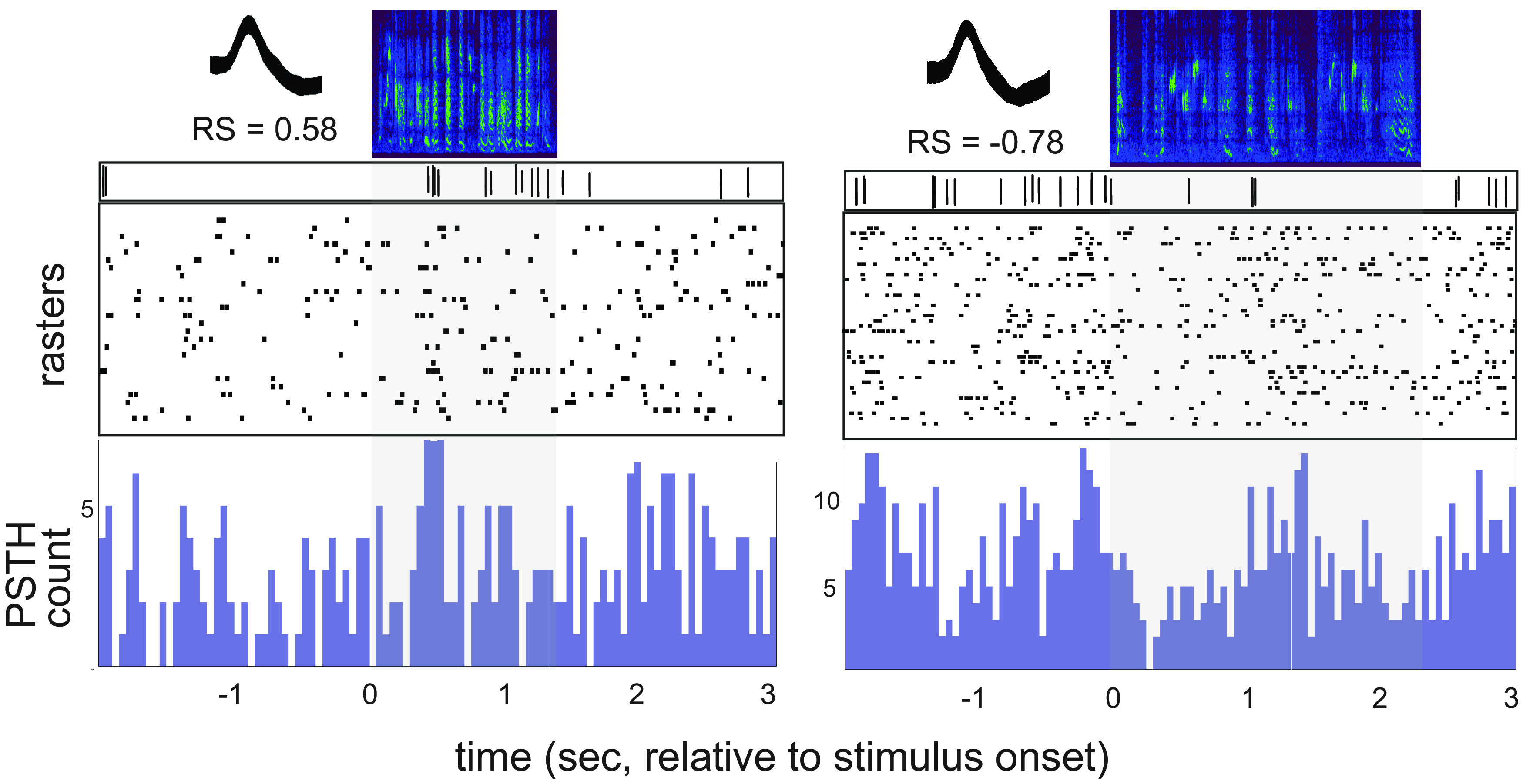
Vocal learning in songbirds is mediated by a highly localized system of interconnected forebrain regions, including recurrent loops that traverse the cortex, basal ganglia, and thalamus. This brain-behavior system provides a powerful model for elucidating mechanisms of vocal learning, with implications for learning speech in human infants, as well as for advancing our understanding of skill learning in general. A long history of experiments in this area has tested neural responses to playback of different song stimuli in anesthetized birds at different stages of vocal development. These studies have demonstrated selectivity for different song types that provide neural signatures of learning. In contrast to the ease of obtaining responses to song playback in anesthetized birds, song-evoked responses in awake birds are greatly reduced or absent, indicating that behavioral state is an important determinant of neural responsivity. Song-evoked responses can be elicited during sleep as well as anesthesia, and the selectivity of responses to song playback in adult birds is highly similar between anesthetized and sleeping states, encouraging the idea that anesthesia and sleep are similar. In contrast to that idea, we report evidence that cortical responses to song playback in juvenile zebra finches (Taeniopygia guttata) differ greatly between sleep and urethane anesthesia. This finding indicates that behavioral states differ in sleep versus anesthesia and raises questions about relationships between developmental changes in sleep activity, selectivity for different song types, and the neural substrate for vocal learning.

Keywords: anesthesia; sensory gating; sleep; songbird; spiking activity; vocal learning.


Figures

Figure 1.
Figure 1.
A simplified schematic of cortico-basal ganglia circuits that mediate vocal learning and a timeline of vocal development. Top, The cortical nucleus LMAN comprises CORE (gray) and SHELL (red) subregions which form parallel recurrent loops through the basal ganglia and dorsal thalamus. LMAN-SHELL also forms a trans-cortical loop via AId that converges with basal ganglia loops in the same dorsal thalamic zone. A transient projection from LMAN-CORE to AId is present only in juvenile birds and creates a site of integration between CORE and SHELL pathways in AId during early sensorimotor learning (denoted by dotted line). The dorsal thalamic zone feeds back to LMAN and feeds forward to HVC via medial MAN (latter pathway not shown for clarity). A specific region of the basal ganglia known as area X is dedicated to functions for vocal learning and includes both striatal and pallidal cells. RA: robust nucleus of the arcopallium; AId: dorsal intermediate arcopallium; HVC: high vocal center; LMAN: lateral magnocellular nucleus of the anterior nidopallium. Bottom, Zebra finches fledge from the nest ∼20 dph and are still reliant on parents to feed and preen them; juvenile males memorize the song of their biological father in the period from ∼20 to 35 dph. They begin to produce their first song-related vocalizations (babbling) ∼35 dph, and gradually refine their vocal motor output until they achieve a stable imitation of their memorized tutor song ∼80–90 dph; they produce a highly stereotyped song throughout adulthood.
Figure 2.
Figure 2.
Proportion of significant responses to each song stimulus in CORE (gray) versus SHELL (red) neurons. Top, Proportions of excited and suppressed responses to playback of each song type (see Table 1). *p = 0.04, **p = 0.01, ***p = 0.003. Bottom, Proportions of suppressed responses to each song type. *p = 0.03, **p = 0.01, ***p = 0.006. AdlCon, adult conspecific song; JuvCon, juvenile conspecific song; TUT, tutor song; OWN, bird’s own song. n = 44 responses in 35 CORE neurons; n = 63 responses in 49 SHELL neurons.
Figure 3.
Figure 3.
Single neurons were selectively tuned in both CORE and SHELL. Left, Proportions of neurons that responded to different numbers of songs out of the four song types played; most neurons (∼75%) responded to only one song stimulus in both CORE and SHELL (green), some neurons (20–26%) responded to two songs (yellow), and two neurons (4%) in SHELL responded to three songs. Right, Each row corresponds to one neuron, indicating the song stimuli to which each neuron responded (n = 44 responses in 35 CORE neurons; n = 63 responses in 49 SHELL neurons). Rows are ordered according to whether each neuron responded to one, two, or three songs (colors corresponding to those on the left). Columns depict responses to each song type, with darker shading indicating suppressed responses and lighter shading indicating excited responses; unshaded boxes depict nonsignificant responses.
Figure 4.
Figure 4.
Firing rates and standardized response strengths in SHELL and CORE neurons. Left panel, Top shows spontaneous firing rates (averages ± SEM) during periods marked as sleeping versus nonsleeping; bottom shows burst fractions (percent of ISIs < 5 ms). One CORE neuron and two SHELL neurons were omitted from the graph of burst fractions since they were outliers (but were included in statistical analyses). **p < 0.005, ***p < 0.0001. Right panel, All graphs depict absolute values (ABS) of standardized response strengths as a function of song type. Top, Response strengths (including both excitatory and suppressed responses, both significant and nonsignificant) for all CORE and SHELL neurons (n = 35 CORE, n = 49 SHELL). * indicates main effect between songs in CORE, p = 0.039. Middle, Response strengths for the subset of CORE and SHELL neurons that showed a significant response to JuvCon song (n = 19 CORE, n = 21 SHELL). Bottom, Response strengths for the subset of CORE and SHELL neurons that showed a significant response to OWN song (n = 11 CORE, n = 21 SHELL). Box-and-whisker plots depict medians and first and third quartiles; whiskers in right panel indicate minimum and maximum values, and circles represent individual data points.
Figure 5.
Figure 5.
Average response strengths to each song stimulus for suppressed versus excited responses. Top panels, Suppressed responses including all response strengths less than zero (left) and excited responses including all response strengths greater than zero (right). Bottom panels, Significant responses (firing rates were significantly different from baseline), plotted as in top panels. Numbers just above/below each bar represent ns (ns for significant responses are also given in Table 1). Averages ± SEM.
Figure 6.
Figure 6.
Two example single units during playback of JuvCon song. Units are from the same bird at 44 dph. Left, CORE neuron that showed excitation to JuvCon. Right, SHELL neuron that showed suppression to JuvCon. For each panel, top half shows song spectrograms and raw traces of single-unit activity; bottom half shows rasters and PSTHs. Overlaid waveforms shown in inset at top left; RS, mean response strength.
Figure 7.
Figure 7.
CORE and SHELL neurons were equally selective for JuvCon song. Each panel shows cumulative distribution functions of selectivity scores for JuvCon song compared with AdlCon (top), TUT (middle), and OWN (bottom; n = 13 CORE, n = 19 SHELL). Positive difference scores indicate a preference for JuvCon song over comparison songs, and show that both CORE (gray) and SHELL (red) neurons preferred JuvCon song over comparison songs to the same extent.

Similar articles

References

    1. Achiro JM, Bottjer SW (2013) Neural representation of a target auditory memory in a cortico-basal ganglia pathway. J Neurosci 33:14475–14488. 10.1523/JNEUROSCI.0710-13.2013 - DOI - PMC - PubMed
    1. Achiro JM, Shen J, Bottjer SW (2017) Neural activity in cortico-basal ganglia circuits of juvenile songbirds encodes performance during goal-directed learning. Elife 6:e26973. 10.7554/eLife.26973 - DOI - PMC - PubMed
    1. Adret P, Meliza CD, Margoliash D (2012) Song tutoring in presinging zebra finch juveniles biases a small population of higher-order song-selective neurons toward the tutor song. J Neurophysiol 108:1977–1987. 10.1152/jn.00905.2011 - DOI - PMC - PubMed
    1. Alexander GE, Crutcher MD (1990) Functional architecture of basal ganglia circuits: neural substrates of parallel processing. Trends Neurosci 13:266–271. 10.1016/0166-2236(90)90107-l - DOI - PubMed
    1. Alvarez-Buylla A, Theelen M, Nottebohm F (1988) Birth of projection neurons in the higher vocal center of the canary forebrain before, during and after song learning. Proc Natl Acad Sci U S A 85:8722–8726. 10.1073/pnas.85.22.8722 - DOI - PMC - PubMed

LinkOut - more resources