

Comparison of the Interference Effects on Somatosensory Evoked Potential from Tonic, Burst, and High-dose Spinal Cord Stimulations
- PMID: 35545502
- PMCID: PMC9357458
- DOI: 10.2176/jns-nmc.2021-0298
Comparison of the Interference Effects on Somatosensory Evoked Potential from Tonic, Burst, and High-dose Spinal Cord Stimulations
Abstract
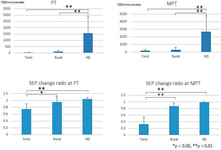
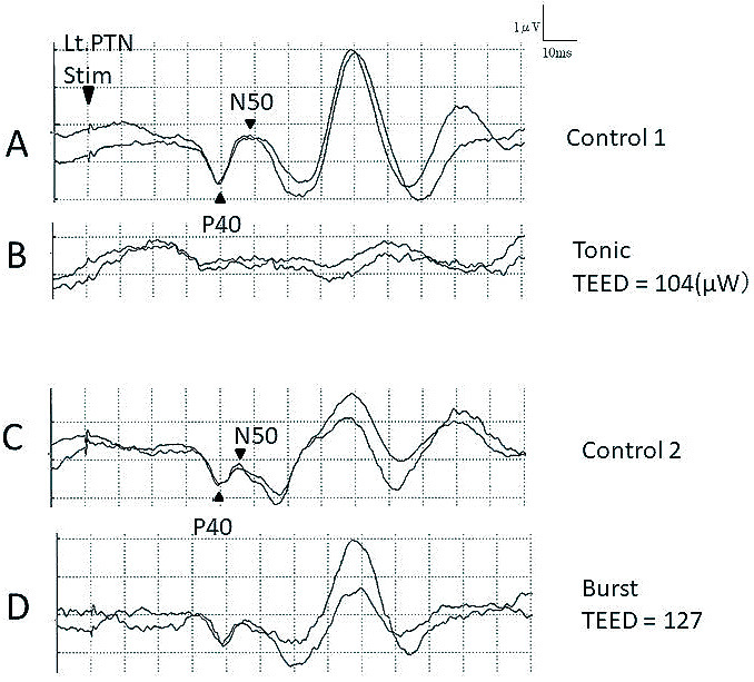
Spinal cord stimulations have been used widely to treat intractable neuropathic pain. The conventional spinal cord stimulation paradigm, the "tonic" type, suppresses excessive activation of wide dynamic range neurons in the dorsal horn via the collateral branch from the dorsal column. Therefore, preserved dorsal column function is an important prerequisite for tonic spinal cord stimulations. A tonic spinal cord stimulation requires eliciting paresthesia in the painful area due to stimulation of the dorsal column and dorsal root. Recent spinal cord stimulation paradigms, including burst and high-dose, are set below the paresthesia threshold and are proposed to have different pain reduction mechanisms. We conducted an interference study of these different stimulation paradigms on the somatosensory evoked potential (SEP) to investigate differences in the sites of action between tonic and new spinal cord stimulations. We recorded posterior tibial nerve-stimulated SEP in seven patients with neuropathic pain during tonic, burst, and high-dose stimulations. The total electrical energy delivered was calculated during SEP-spinal cord stimulation interference studies. High-dose stimulations could not reduce the SEP amplitude despite higher energy delivery than tonic stimulation. Burst stimulation with an energy similar to the tonic stimulation could not reduce SEP amplitude as tonic stimulation. The study results suggested different sites of action and effects on the spinal cord between the conventional tonic and burst or high-dose spinal cord stimulations.
Keywords: burst stimulation; high-dose stimulation; somatosensory evoked potentials; spinal cord stimulation; tonic stimulation.
Conflict of interest statement
All authors have no conflict of interest in relation to this manuscript.
Figures


Similar articles
-
Burst spinal cord stimulation for limb and back pain.World Neurosurg. 2013 Nov;80(5):642-649.e1. doi: 10.1016/j.wneu.2013.01.040. Epub 2013 Jan 12. World Neurosurg. 2013. PMID: 23321375 Clinical Trial.
-
Pulse Intensity Effects of Burst and Tonic Spinal Cord Stimulation on Neural Responses to Brushing in Patients With Neuropathic Pain.Neuromodulation. 2023 Jul;26(5):975-987. doi: 10.1016/j.neurom.2022.11.001. Epub 2022 Dec 1. Neuromodulation. 2023. PMID: 36464560
-
Spinal cord stimulation for intractable pain evaluated by a collision study using somatosensory evoked potentials: a preliminary report.Neuromodulation. 2014 Dec;17(8):746-52; discussion 752. doi: 10.1111/ner.12205. Epub 2014 Jun 19. Neuromodulation. 2014. PMID: 24945895
-
Mechanisms and mode of action of spinal cord stimulation in chronic neuropathic pain.Postgrad Med. 2020 Nov;132(sup3):17-21. doi: 10.1080/00325481.2020.1769393. Epub 2020 May 22. Postgrad Med. 2020. PMID: 32403963 Review.
-
Tonic, Burst, High-Density, and 10-kHz High-Frequency Spinal Cord Stimulation: Efficiency and Patients' Preferences in a Failed Back Surgery Syndrome Predominant Population. Review of Literature.World Neurosurg. 2020 Dec;144:e331-e340. doi: 10.1016/j.wneu.2020.08.128. Epub 2020 Sep 2. World Neurosurg. 2020. PMID: 32889188 Review.
Cited by
-
Unaltered Responses of Distal Motor Neurons to Non-Targeted Thoracic Spinal Cord Stimulation in Chronic Pain Patients.Pain Ther. 2024 Dec;13(6):1645-1658. doi: 10.1007/s40122-024-00670-x. Epub 2024 Oct 18. Pain Ther. 2024. PMID: 39424774 Free PMC article.
References
-
- Guan Y, Wacnik PW, Yang F, et al. : Spinal cord stimulation-induced analgesia: Electrical stimulation of dorsal column and dorsal roots attenuates dorsal horn neuronal excitability in neuropathic rats. Anesthesiology 113: 1392-1405, 2010 - PubMed
-
- Yakhnitsa V, Linderoth B, Meyerson BA: Spinal cord stimulation attenuates dorsal horn neuronal hyperexcitability in a rat model of mononeuropathy. Pain 79: 223-233, 1999 - PubMed
-
- Wallin J, Fiskå A, Tjølsen A, Linderoth B, Hole K: Spinal cord stimulation inhibits long-term potentiation of spinal wide dynamic range neurons. Brain Res 973: 39-43, 2003 - PubMed