. 2022 May 11;12(1):7775.
doi: 10.1038/s41598-022-11755-y.

Effect of ambient lighting on frequency dependence in transcranial electrical stimulation-induced phosphenes

Affiliations

Effect of ambient lighting on frequency dependence in transcranial electrical stimulation-induced phosphenes

Ian Evans et al. Sci Rep. .

Abstract

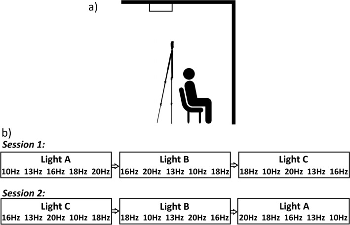
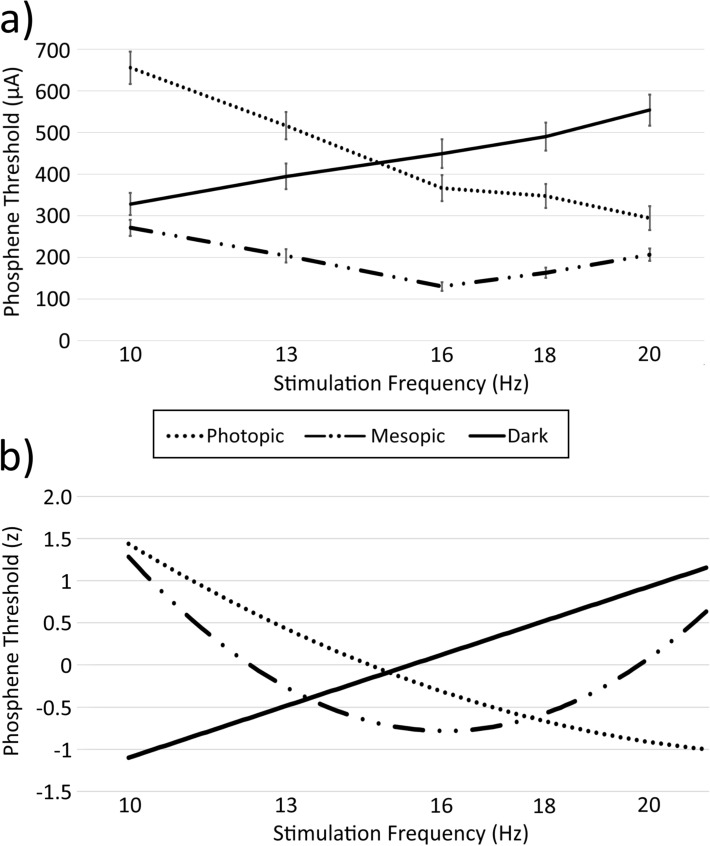
Inconsistencies have been found in the relationship between ambient lighting conditions and frequency-dependence in transcranial electric stimulation (tES) induced phosphenes. Using a within-subjects design across lighting condition (dark, mesopic [dim], photopic [bright]) and tES stimulation frequency (10, 13, 16, 18, 20 Hz), this study determined phosphene detection thresholds in 24 subjects receiving tES using an FPz-Cz montage. Minima phosphene thresholds were found at 16 Hz in mesopic, 10 Hz in dark and 20 Hz in photopic lighting conditions, with these thresholds being substantially lower for mesopic than both dark (60% reduction) and photopic (56% reduction), conditions. Further, whereas the phosphene threshold-stimulation frequency relation increased with frequency in the dark and decreased with frequency in the photopic conditions, in the mesopic condition it followed the dark condition relation from 10 to 16 Hz, and photopic condition relation from 16 to 20 Hz. The results clearly demonstrate that ambient lighting is an important factor in the detection of tES-induced phosphenes, and that mesopic conditions are most suitable for obtaining overall phosphene thresholds.


Conflict of interest statement

The authors declare no competing interests.

Figures

Figure 1
Figure 1
(a) Positioning of lighting and test subject. The test subject was seated so that the front wall filled their entire field of view (no parts of either side wall were visible). The light stand was positioned so that no shadows were visible in the subject’s field of view. The photopic lighting condition was achieved by activating the fixed fluorescent ceiling light with no light stand used, while all other lighting conditions (the mesopic condition and the brief periods requiring light in the dark condition) were achieved using the light stand only. (b) Example sequence of the block design across both sessions. For the first session, each lighting condition block was presented in a random order, as were the frequencies within it. In order to account for potential within-subject order effects due to possible light adaptation, in the second session the order of the lighting blocks was reversed and the frequencies within those blocks were reversed.
Figure 2
Figure 2
(a) Phosphene thresholds and standard errors for each ambient lighting condition at each frequency tested (10, 13, 16, 18 and 20 Hz). (b) Regression-based estimates of normalised phosphene thresholds, as a function of stimulation frequency, for each ambient lighting condition.

References

    1. Buzsáki G, Draguhn A. Neuronal oscillations in cortical networks. Science. 2004;304:1926–1929. doi: 10.1126/science.1099745. - DOI - PubMed
    1. Herrmann CS, Fründ I, Lenz D. Human gamma-band activity: A review on cognitive and behavioral correlates and network models. Neurosci. Biobehav. Rev. 2010;34:981–992. doi: 10.1016/j.neubiorev.2009.09.001. - DOI - PubMed
    1. Bosman CA, Lansink CS, Pennartz CM. Functions of gamma-band synchronization in cognition: From single circuits to functional diversity across cortical and subcortical systems. Eur. J. Neurosci. 2014;39:1982–1999. doi: 10.1111/ejn.12606. - DOI - PubMed
    1. Schwiedrzick C. Retina or visual cortex? The site of phosphene induction by transcranial alternating current stimulation. Front. Integr. Neurosci. 2009;3(6):1–2. - PMC - PubMed
    1. Schutter DJ, Hortensius R. Retinal origin of phosphenes to transcranial alternating current stimulation. Clin. Neurophysiol. 2010;121:1080–1084. doi: 10.1016/j.clinph.2009.10.038. - DOI - PubMed

Publication types