. 2022 May;605(7910):527-531.
doi: 10.1038/s41586-022-04717-x. Epub 2022 May 11.

Group A Streptococcus induces GSDMA-dependent pyroptosis in keratinocytes

Affiliations

Group A Streptococcus induces GSDMA-dependent pyroptosis in keratinocytes

Doris L LaRock et al. Nature. 2022 May.

Abstract

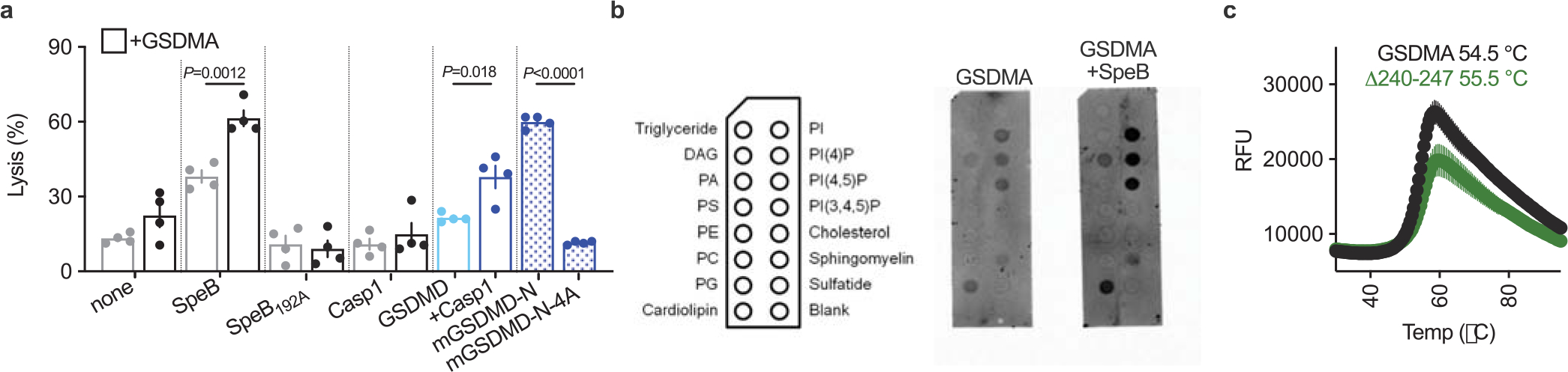
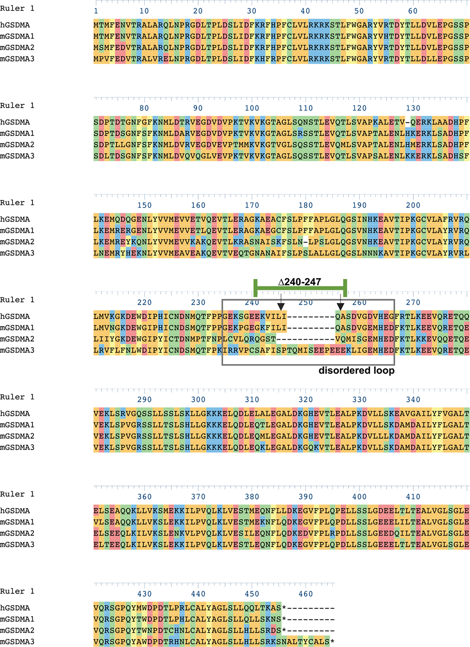
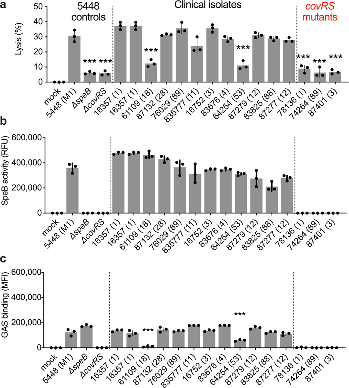
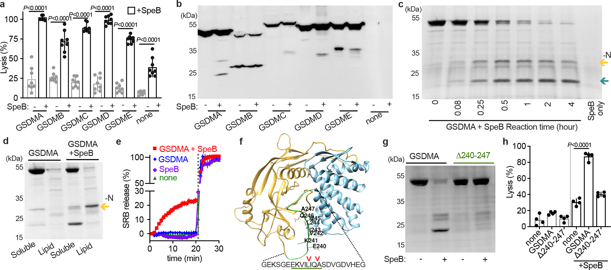
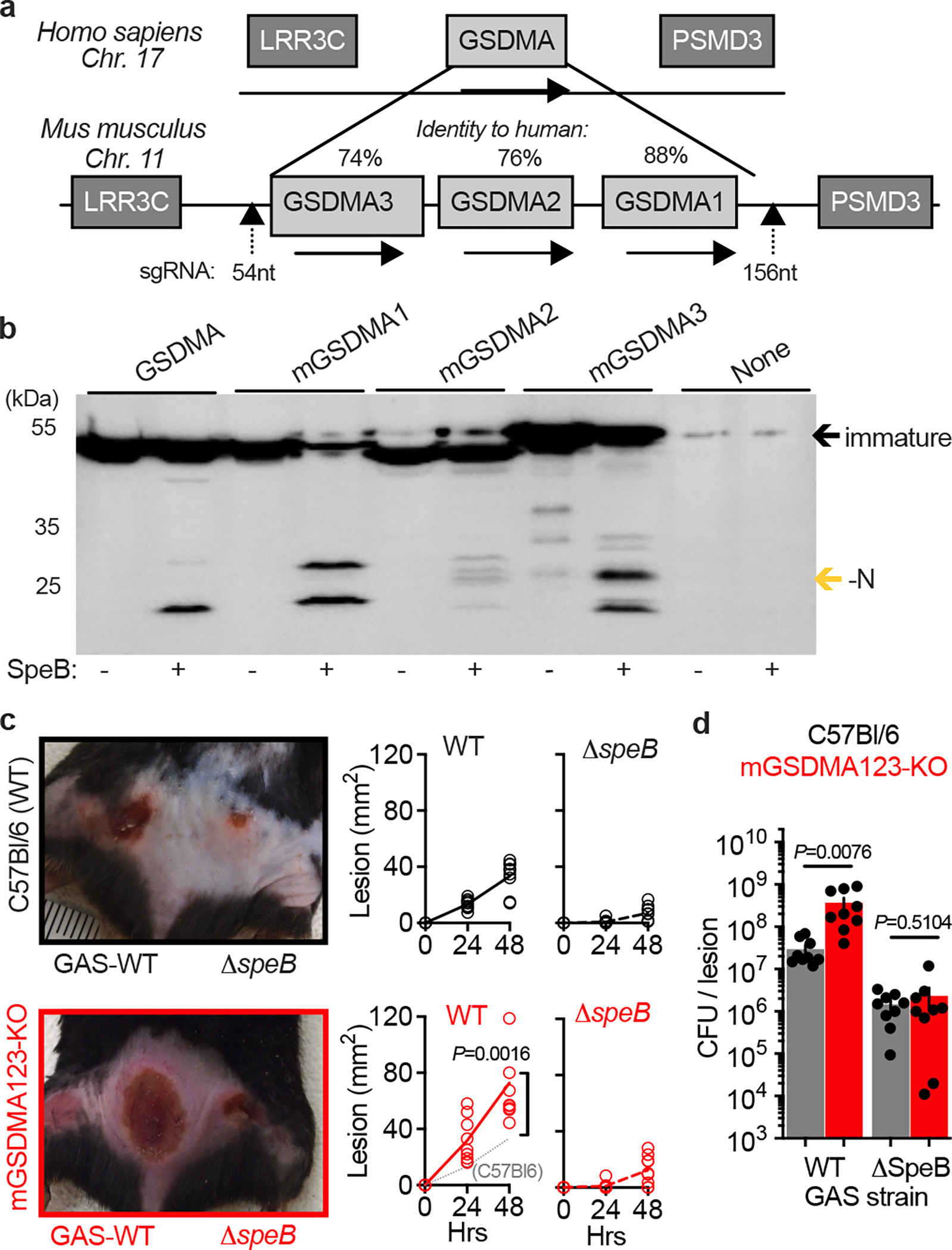
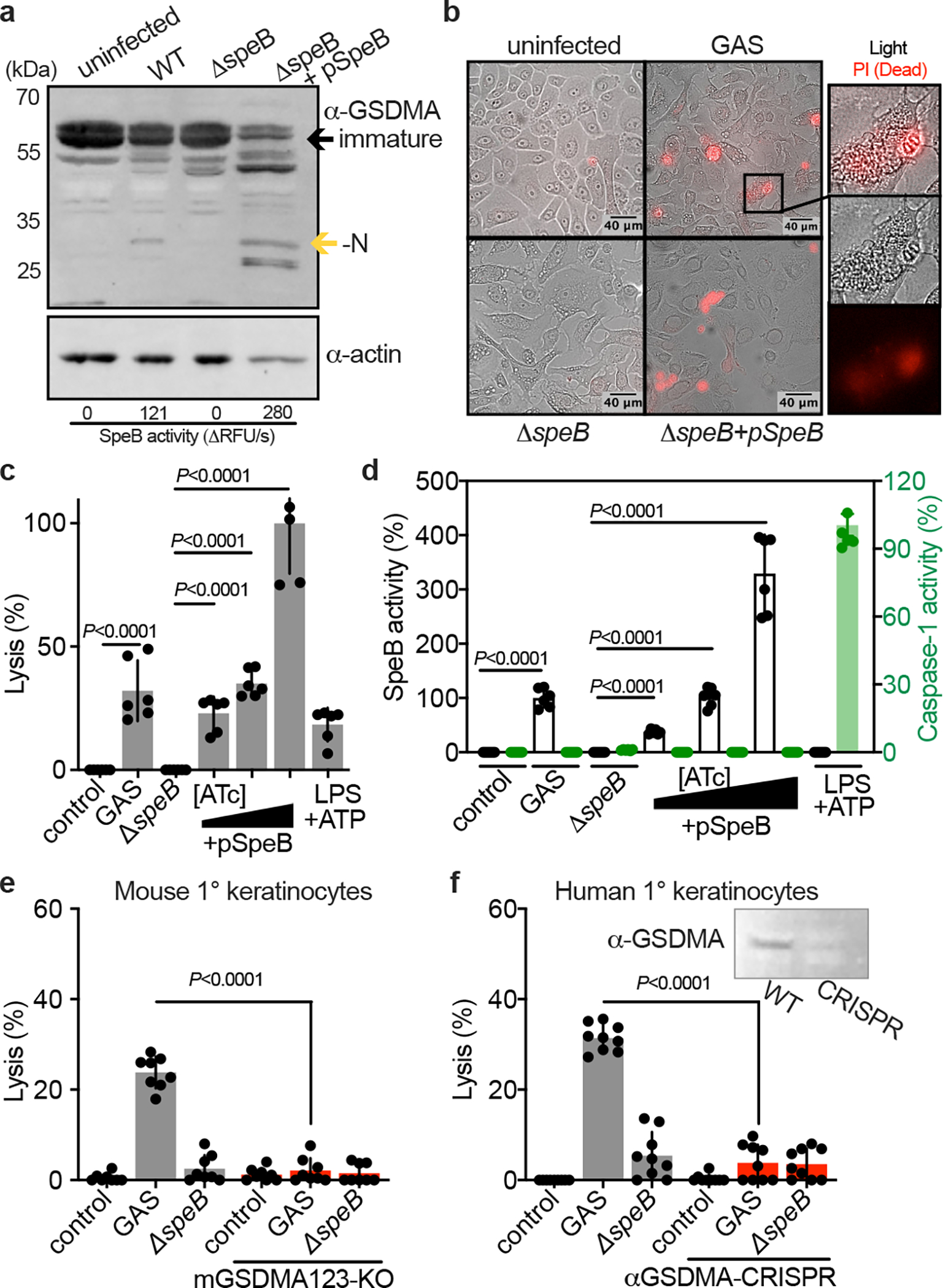
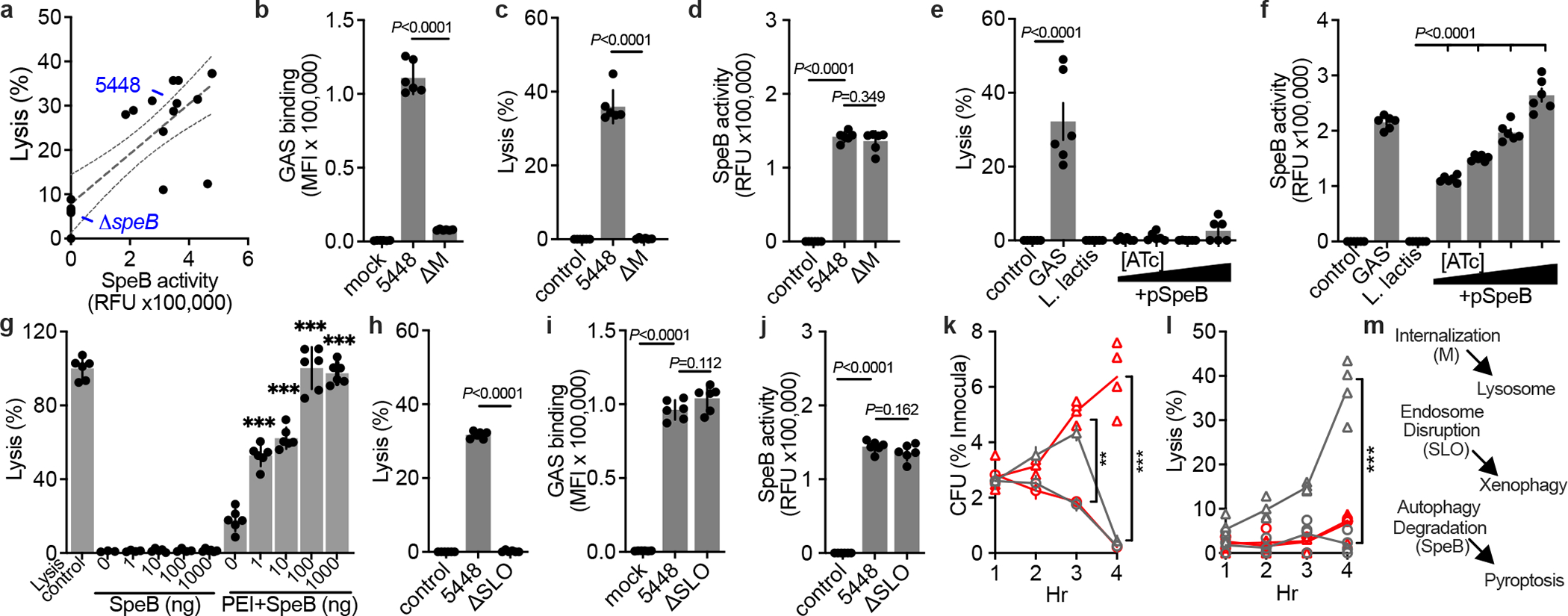
Gasdermins (GSDMs) are a family of pore-forming effectors that permeabilize the cell membrane during the cell death program pyroptosis1. GSDMs are activated by proteolytic removal of autoinhibitory carboxy-terminal domains, typically by caspase regulators1-9. However, no activator is known for one member of this family, GSDMA. Here we show that the major human pathogen group A Streptococcus (GAS) secretes a protease virulence factor, SpeB, that induces GSDMA-dependent pyroptosis. SpeB cleavage of GSDMA releases an active amino-terminal fragment that can insert into membranes to form lytic pores. GSDMA is primarily expressed in the skin10, and keratinocytes infected with SpeB-expressing GAS die of GSDMA-dependent pyroptosis. Mice have three homologues of human GSDMA, and triple-knockout mice are more susceptible to invasive infection by a pandemic hypervirulent M1T1 clone of GAS. These results indicate that GSDMA is critical in the immune defence against invasive skin infections by GAS. Furthermore, they show that GSDMs can act independently of host regulators as direct sensors of exogenous proteases. As SpeB is essential for tissue invasion and survival within skin cells, these results suggest that GSDMA can act akin to a guard protein that directly detects concerning virulence activities of microorganisms that present a severe infectious threat.


Conflict of interest statement

Competing Interests D.L.L. and C.N.L. are named inventors on a patent application that describes GSDMA activities.

Figures

Extended Data Fig. 1 |
Extended Data Fig. 1 |. SpeB cleaves GSDMA.
a, Cytotoxicity of HEK293Ts transfected with human GSDMs ± SpeB. Data are the mean±s.d. of 4 technical replicates. P values were calculated by two-tailed Student’s t-test. b, Membranes spotted with lipids were incubated with indicated proteins and binding was assessed by immunoblot for GSDMA. c, Thermal melt analysis of GSDMA and GSDMAΔ240-247 with melting temperatures indicated. (a-c) data are representative of three independent experiments.
Extended Data Fig 2. |
Extended Data Fig 2. |. Alignment of human and mouse GSDMAs.
Human GSDMA (Q96QA5), mouse GSDMA_1 (Q9EST1), Mouse GSDMA_2 (Q32M21), mouse GSDMA_3 (Q5Y4Y6) aligned with Clustal Omega algorithm in DNASTAR. Disordered solvent accessible loop (gray box). Inverted arrows identify cleavage sites of SpeB on hGSDMA. Residues 240–247 of hGSDMA are identified (green bar).
Extended Data Fig 3. |
Extended Data Fig 3. |. Lytic activity, SpeB production, and binding of GAS strains.
a,b, Keratinocytes were infected (MOI=100) with GAS 5448, isogenic mutant controls, or clinical isolates for 4 h and (a) lysis measured by LDH release and (b) SpeB activity measured using specific substrate Mca-IFFDTWDNE-Lys-Dnp. c, GAS labeled with Bocillin was incubated with keratinocytes (MOI=100) 1 h, washed, and adherence measured by fluorescence. Data are representative of three independent experiments with 3 technical replicates and are presented as the mean±s.d. P values were calculated by two-way ANOVA compared to 5448 (M1) control; P<0.0001.
Fig. 1 |
Fig. 1 |. SpeB cleaves GSDMA.
a, Cytotoxicity of each human GSDMs transfected ±SpeB in HEK293Ts. Data are the mean±s.d. of 8 technical replicates. b, Lysates of GSDM-transfected HEK293Ts incubated with SpeB and analysed by immunoblot. c, SDS–PAGE of recombinant human GSDMA cleavage by SpeB over time. GSDMA-N (gold arrow), GSDMA-C (teal arrow). d, PC:PE:cardiolipin liposome binding of GSDMA ±SpeB analysed by SDS–PAGE. GSDMA-N (gold arrow). e, Liposome leakage monitored by Sulforhodamine B (SRB) fluorescence on incubation with GSDMA ±SpeB. Detergent was added after 21 min (dotted line). f, hGSDMA model (Alphafold:Q96QA5) with SpeB cleavage sites identified by Edman sequencing indicated by arrows; GSDMAΔ240-247 deletion underlined in green. g, Cleavage of recombinant GSDMA or GSDMAΔ240-247 ±SpeB was analysed by SDS-PAGE. h, Cytotoxicity of HEK293Ts transfected with GSDMA or GSDMAΔ240-247 ±SpeB. Data are the mean±s.d. of 4 technical replicates. P values were calculated by two-tailed unpaired Student’s t-test (a,h) Data (a-h) are representative of three independent experiments. For gel source data, see Supplementary Figure 1.
Fig. 2 |
Fig. 2 |. GSDMA protects mice against severe GAS skin infection
a, GSDMA gene arrangement in mice, with identity to human GSDMA and site of CRISPR knockout indicated. b, Lysates of HEK293Ts transfected with indicated plasmids were incubated with purified SpeB and analysed by immunoblot for Flag-tagged GSDMs (GSDM-N indicated by gold arrow). One of three repeats is shown. For gel source data, see Supplementary Figure 1. c,d, Wild-type (C57Bl/6) and GSDMA-deficient (mGSDMA123-KO) mice were inoculated intradermally with 108 CFU GAS 5448 or ΔspeB and (c) lesion area measured and compared to GAS-WT in C57Bl/6 mice (dotted line). Representative images 48 h post-infection are shown. (d) GAS CFU enumeration in the same mice 48 h post-infection. Data (c-d) are pooled from two independent experiments and are the mean±s.d. (n=9). P values were calculated by two-tailed unpaired Student’s t-test.
Fig. 3 |
Fig. 3 |. GAS induce SpeB-dependent keratinocyte pyroptosis
a, Keratinocytes were infected with GAS (MOI=100) for 24 h and GSDMA cleavage was detected by immunoblot. SpeB activity reported as initial kinetic velocity was measured using specific substrate Mca-IFFDTWDNE-Lys-Dnp. b, Immunofluorescent microscopy images of keratinocyte permeabilization to propidium iodide (PI; red) during GAS infection (MOI=100), 2 h. (a,b) Data are representative of three independent experiments. c,d, Human keratinocytes infected 4 h with GAS (MOI=100), ΔspeB, and pSpeB complemented ΔspeB, inducible with 0.1, 1, or 10 ng/ml Anhydrotetracycline (ATc). (c) Lysis measured by LDH release and (d) SpeB and Caspase activity measured using specific substrates Mca-IFFDTWDNE-Lys-Dnp and YVAD-pNA. Cells were treated 18 h with LPS 100 ng/ml and 2 h ATP 5 mM as a caspase-1 control. Data are the mean±s.d. of 6 technical replicates. e,f, Keratinocytes were infected for 4 h with GAS or ΔspeB (MOI=100), treated with gentamycin at 1 h, and lysis measured by LDH release. Data are the mean±s.d. of 9 technical replicates. (e) Keratinocytes were isolated from C57Bl/6 or mGSDMA123-KO mice. (f) Human keratinocytes transfected with GSDMA-targeting sgRNA:Cas9 complex or mock-treated 72 h before infection. Efficacy was evaluated by GSDMA immunoblot. (c,d,e,f) P values were calculated by two-tailed unpaired Student’s t-test and are representative of three independent experiments. For gel source data, see Supplementary Figure 1.
Fig. 4 |
Fig. 4 |. GSDMA activation requires cell contact and restricts iGAS
a-f,h-j. Human keratinocytes were infected MOI=100, treated with gentamycin at 1 h, then (a,c,e,h), lysis measured by LDH release after 4 h, (a,d,f,j) SpeB activity measured using specific substrate Mca-IFFDTWDNE-Lys-Dnp after 4 h, and (b,i) Bocillin-labeled GAS adherence after PBS wash was measured by fluorescence after 1 h incubation. (f) 0, 0.1, 1, or 10 ng/mL ATc was included for SpeB induction. g, Keratinocyte lysis measured by LDH release 2 h after treatment with SpeB dilutions (P=0.307; 0.082; 0.330; 0.0243) or packaged within PEI liposomes (all P<0.0001). (a-j) Data are representative of three independent experiments and presented as mean±s.d. of 6 technical replicates. k,l, Human normal (gray) or GSDMA-KO (red) keratinocytes were infected (MOI=100) with JRS4 (circles) or JRS4+pSpeB (triangles) and (k) intracellular CFU and (l) lysis evaluated by LDH release. JRS4+pSpeB has a growth advantage at 3 h (P=0.0004) lost at 4 h, concurrent with cell lysis (P<0.0001). The growth advantage is sustained in lysis-defective GSDMA-KO keratinocytes(P<0.0001). (k,l) Data are representative of three independent experiments and presented as mean±s.d. of 4 technical replicates. (b-l) P values were calculated by two-tailed unpaired Student’s t-test; **P<0.001,***P<0.0001. m, Proposed model of iGAS virulence factor activities and their detection by host cells.

References

    1. Broz P, Pelegrín P & Shao F The gasdermins, a protein family executing cell death and inflammation. Nat. Rev. Immunol. 20, 143–157 (2019) - PubMed
    1. Zhou Z et al. Granzyme A from cytotoxic lymphocytes cleaves GSDMB to trigger pyroptosis in target cells. Science 368, eaaz7548 (2020). - PubMed
    1. Aglietti RA et al. GsdmD p30 elicited by caspase-11 during pyroptosis forms pores in membranes. Proc. Natl. Acad. Sci. 113, 7858–7863 (2016) - PMC - PubMed
    1. Ding J et al. Pore-forming activity and structural autoinhibition of the gasdermin family. Nature 538, 111–116 (2016) - PubMed
    1. Liu X et al. Inflammasome-activated gasdermin D causes pyroptosis by forming membrane pores. Nature 535, 153–158 (2016). - PMC - PubMed