Digital Pathology and Artificial Intelligence Applications in Pathology
- PMID: 35545826
- PMCID: PMC9098984
- DOI: 10.14791/btrt.2021.0032
Digital Pathology and Artificial Intelligence Applications in Pathology
Abstract
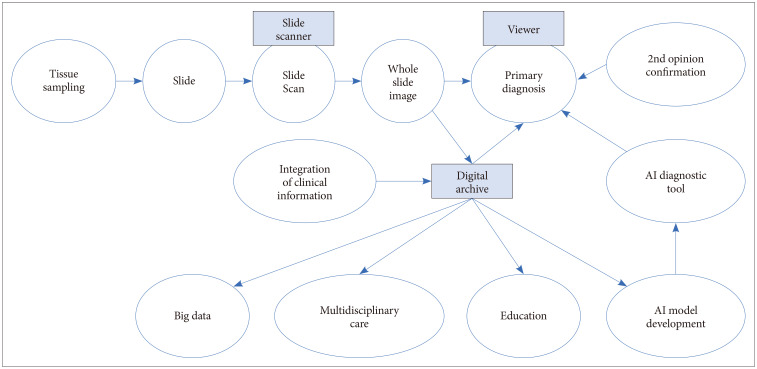
Digital pathology is revolutionizing pathology. The introduction of digital pathology made it possible to comprehensively change the pathology diagnosis workflow, apply and develop pathological artificial intelligence (AI) models, generate pathological big data, and perform telepathology. AI algorithms, including machine learning and deep learning, are used for the detection, segmentation, registration, processing, and classification of digitized pathological images. Pathological AI algorithms can be helpfully utilized for diagnostic screening, morphometric analysis of biomarkers, the discovery of new meanings of prognosis and therapeutic response in pathological images, and improvement of diagnostic efficiency. In order to develop a successful pathological AI model, it is necessary to consider the selection of a suitable type of image for a subject, utilization of big data repositories, the setting of an effective annotation strategy, image standardization, and color normalization. This review will elaborate on the advantages and perspectives of digital pathology, AI-based approaches, the applications in pathology, and considerations and challenges in the development of pathological AI models.
Keywords: Artificial intelligence; Deep learning; Digital technology; Pathology; Workflow.
Copyright © 2022 The Korean Brain Tumor Society, The Korean Society for Neuro-Oncology, and The Korean Society for Pediatric Neuro-Oncology.
Conflict of interest statement
The author has no potential conflicts of interest to disclose.
Figures