Peripheral T-Cell Lymphoma Presenting as a Scalp Mass
- PMID: 35545831
- PMCID: PMC9098978
- DOI: 10.14791/btrt.2022.0004
Peripheral T-Cell Lymphoma Presenting as a Scalp Mass
Abstract
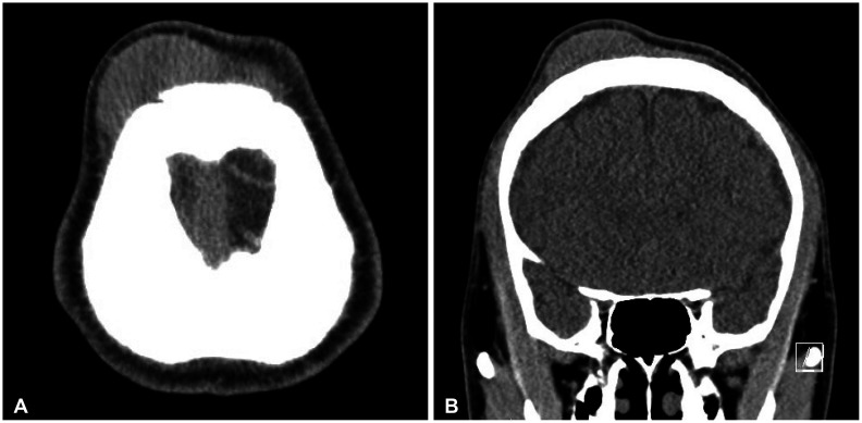
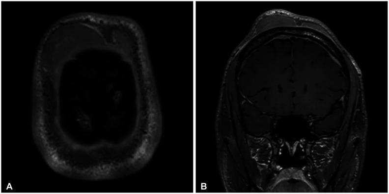
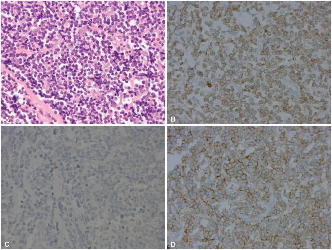
Peripheral scalp T-cell lymphoma is a very rare disease. We report a case of a 22-year-old man who presented an indolent large scalp mass in the right frontal scalp region. The patient's physical examination demonstrated no palpable mass in the chest, abdomen, and extremities. The brain CT revealed a high-density large scalp mass of the subgaleal layer in the right frontal and a small scalp mass of the subgaleal layer in the left frontal. The brain MRI showed multifocal enhancing masses in the bilateral dura, the subgaleal layer of the scalp, and the skull. The patient underwent removal of the tumor found in the right frontal scalp. The histologic diagnosis was peripheral T-cell lymphoma. Bone marrow aspiration showed the involvement of T-cell lymphoma. The patient received chemotherapy with cyclophosphamide, vincristine, doxorubicin, and prednisolone (CHOP protocol) for 3 cycles. The patient was discharged without neurological deficit. The patient showed no evidence of recurrence 15 months after surgery. We report a rare case of peripheral T-cell lymphoma mimicking benign scalp tumors.
Keywords: Lymphoma; Mass; Scalp.
Copyright © 2022 The Korean Brain Tumor Society, The Korean Society for Neuro-Oncology, and The Korean Society for Pediatric Neuro-Oncology.
Conflict of interest statement
The authors have no potential conflicts of interest to disclose.
Figures

Similar articles
-
A case of primary dural lymphoma: diffuse large B-cell type.Turk Neurosurg. 2014;24(5):799-803. doi: 10.5137/1019-5149.JTN.9535-13.2. Turk Neurosurg. 2014. PMID: 25269058
-
Primary diffuse large B-cell lymphomas of the temporoparietal dura mater and scalp without intervening skull bone invasion.Neurol Med Chir (Tokyo). 2010;50(7):595-8. doi: 10.2176/nmc.50.595. Neurol Med Chir (Tokyo). 2010. PMID: 20671390
-
Diffuse large B cell lymphoma of the cranial vault: two case reports.Brain Tumor Pathol. 2015 Oct;32(4):275-80. doi: 10.1007/s10014-015-0225-5. Epub 2015 Jul 16. Brain Tumor Pathol. 2015. PMID: 26177806
-
Primary bilateral adrenal non-Hodgkin's Burkitt-like lymphoma: a rare cause of primary adrenal insufficiency. Case report and literature review.Tumori. 2007 Nov-Dec;93(6):625-30. doi: 10.1177/030089160709300621. Tumori. 2007. PMID: 18338503 Review.
-
Aggressive primary scalp lymphoma mimicking an acute epidural hematoma: Case report and review of the literature.Neurochirurgie. 2022 Oct;68(5):e34-e39. doi: 10.1016/j.neuchi.2022.03.005. Epub 2022 Apr 25. Neurochirurgie. 2022. PMID: 35477013 Review.
Cited by
-
Association of weight loss strategies with all-cause and specific-cause mortality: a prospective cohort study.BMC Public Health. 2024 Aug 16;24(1):2234. doi: 10.1186/s12889-024-19472-z. BMC Public Health. 2024. PMID: 39152410 Free PMC article.
References
-
- Duyndam DA, Biesma DH, van Heesewijk JP. Primary non-Hodgkin’s lymphoma of the cranial vault; MRI features before and after treatment. Clin Radiol. 2002;57:948–950. - PubMed
-
- Batchelor TT. In: Youmans and Winn neurological surgery. 7th ed. Richard Winn H, editor. Philadelphia: Elsevier; 2017. Primary central nervous sysytem lymphomas; pp. 1085–1090.
-
- Ikumi N, Fujita H, Terui T, Takahashi H, Miura K, Hatta Y, et al. Aggressive CD4–CD8–CD45RA+CCR10– primary cutaneous peripheral T-cell lymphoma, not otherwise specified: a case report. Acta Derm Venereol. 2019;99:1176–1177. - PubMed
Publication types
Grants and funding
LinkOut - more resources
Full Text Sources
Research Materials