Rapid-Growing Intracranial Immature Teratoma Presenting Obstructive Hydrocephalus and Abducens Nerve Palsy: A Case Report and Literature Review
- PMID: 35545832
- PMCID: PMC9098977
- DOI: 10.14791/btrt.2022.0005
Rapid-Growing Intracranial Immature Teratoma Presenting Obstructive Hydrocephalus and Abducens Nerve Palsy: A Case Report and Literature Review
Abstract
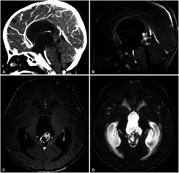
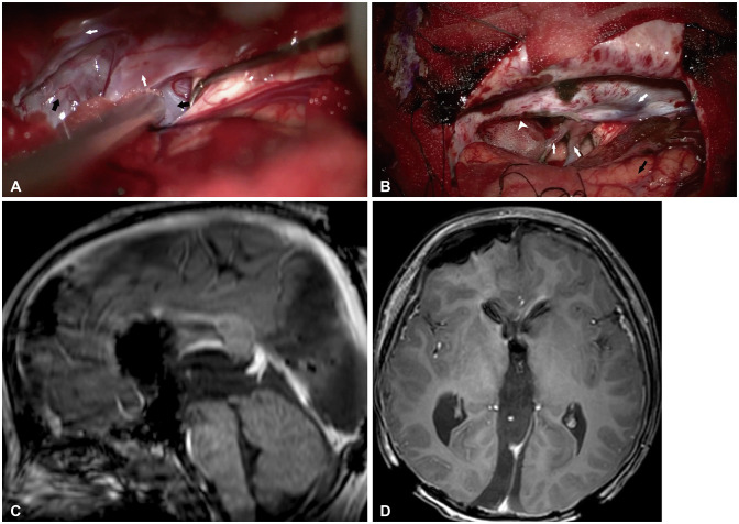
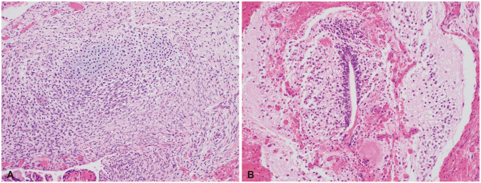
Intracranial immature teratoma is an extremely rare disease with poor prognosis and requires complicated treatment. Owing to the deep midline location of the tumor, total surgical resection of the tumor is challenging. We present our experience with a fast-growing pineal gland immature teratoma in a 4-year-old boy, who presented with obstructive hydrocephalus and abducens nerve palsy, which was treated with total surgical resection of the tumor. In addition, we aimed to determine the appropriate treatment modality for intracranial immature teratomas by reviewing the literature and investigating the prognosis.
Keywords: Brain neoplasms; Hydrocephalus; Immature teratoma.
Copyright © 2022 The Korean Brain Tumor Society, The Korean Society for Neuro-Oncology, and The Korean Society for Pediatric Neuro-Oncology.
Conflict of interest statement
The authors have no potential conflicts of interest to disclose.
Figures


References
-
- Goyal N, Kakkar A, Singh PK, Sharma MC, Chandra PS, Mahapatra AK, et al. Intracranial teratomas in children: a clinicopathological study. Childs Nerv Syst. 2013;29:2035–2042. - PubMed
-
- Georgiu C, Opincariu I, Cebotaru CL, Mirescu ŞC, Stănoiu BP, Domşa TA, et al. Intracranial immature teratoma with a primitive neuroectodermal malignant transformation - case report and review of the literature. Rom J Morphol Embryol. 2016;57:1389–1395. - PubMed
-
- Abdelmuhdi AS, Almazam AE, Dissi NA, Albastaki UM, Pierre-Jerome C. Intracranial teratoma: imaging, intraoperative, and pathologic features: AIRP best cases in radiologic-pathologic correlation. Radiographics. 2017;37:1506–1511. - PubMed
-
- Huang X, Zhang R, Zhou LF. Diagnosis and treatment of intracranial immature teratoma. Pediatr Neurosurg. 2009;45:354–360. - PubMed
-
- Lee YH, Park EK, Park YS, Shim KW, Choi JU, Kim DS. Treatment and outcomes of primary intracranial teratoma. Childs Nerv Syst. 2009;25:1581–1587. - PubMed