. 2022 Aug;28(8):1205-1217.
doi: 10.1111/cns.13853. Epub 2022 May 12.

Partial restoration of spinal cord neural continuity via vascular pedicle hemisected spinal cord transplantation using spinal cord fusion technique

Affiliations

Partial restoration of spinal cord neural continuity via vascular pedicle hemisected spinal cord transplantation using spinal cord fusion technique

Xiaoping Ren et al. CNS Neurosci Ther. 2022 Aug.

Abstract

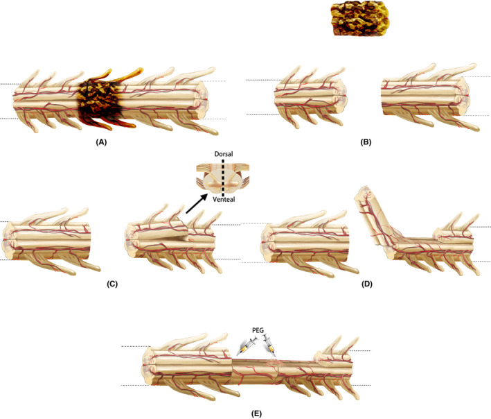
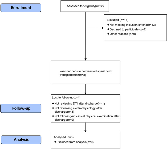
Aims: Our team tested spinal cord fusion (SCF) using the neuroprotective agent polyethylene glycol (PEG) in different animal (mice, rats, and beagles) models with complete spinal cord transection. To further explore the application of SCF for the treatment of paraplegic patients, we developed a new clinical procedure for SCF called vascular pedicle hemisected spinal cord transplantation (vSCT) and tested this procedure in eight paraplegic participants.

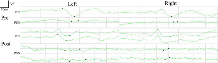
Methods: Eight paraplegic participants (American Spinal Injury Association, ASIA: A) were enrolled and treated with vSCT (PEG was applied to the sites of spinal cord transplantation). Pre- and postoperative pain intensities, neurologic assessments, electrophysiologic monitoring, and neuroimaging examinations were recorded.

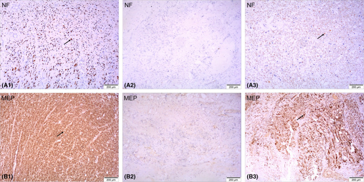
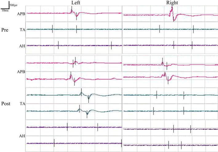
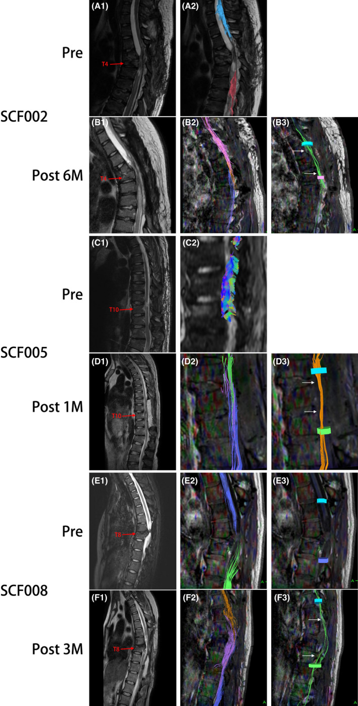
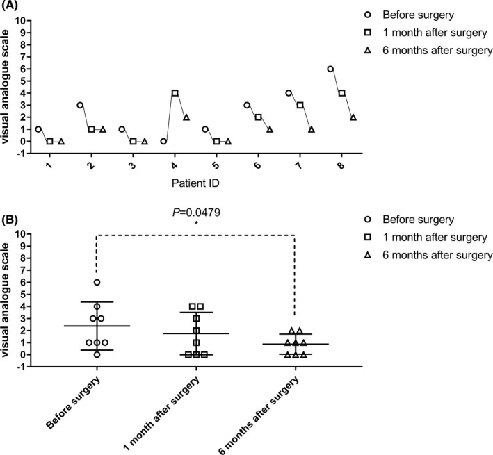
Results: Of the eight paraplegic participants who completed vSCT, objective improvements occurred in motor function for one participant, in electrophysiologic motor-evoked potentials for another participant, in re-establishment of white matter continuity in three participants, in autonomic nerve function in seven participants, and in symptoms of cord central pain for seven participants.

Conclusions: The postoperative recovery of paraplegic participants demonstrated the clinical feasibility and efficacy of vSCT in re-establishing the continuity of spinal nerve fibers. vSCT could provide the anatomic, morphologic, and histologic foundations to potentially restore the motor, sensory, and autonomic nervous functions in paraplegic patients. More future clinical trials are warranted.

Keywords: GEMINI; clinic trial; polyethylene glycol; spinal cord fusion; spinal cord injury.


Conflict of interest statement

The authors declare that they have no conflict of interest.

Figures

FIGURE 1
FIGURE 1
Key surgical steps of vSCT. First, the spinal cord injury area was removed to produce two fresh spinal cord stumps (A, B). Then, based on the effective length of the defect between two fresh spinal stumps, half of the spinal cord tissue with one side of the posterior spinal artery was cut from the distal or proximal spinal cord. The other side of the posterior spinal artery was maintained as the vascular pedicle to supply the blood flow (C). Then half of the spinal cord tissue was transplanted into the spinal cord defect area (D). PEG was topically applied to the two sites of spinal cord transection created after the transplantation to complete the SCF (E)
FIGURE 2
FIGURE 2
CONSORT flow diagram
FIGURE 3
FIGURE 3
Immunohistochemical staining of spinal cord tissue removed during the surgery. NF‐200‐positive axons and MBP‐positive myelin sheaths can be observed at the distal and proximal ends of the tissue (arrows in A1, B1, A3, B3) but not in the center of the tissue (A2, B2)
FIGURE 4
FIGURE 4
Pre‐ and postoperative SSEPs of median nerve (MN) and posterior tibial nerve (PSN) in participant SCF006. Postoperative SSEPs of both lower extremities PSN did not show significant recovery compared with preoperative SSEPs. Pre, preoperatively; Post, postoperatively
FIGURE 5
FIGURE 5
Pre‐ and postoperative MEPs recorded at the abductor pollicis brevis (APB), tibialis anterior (TA), and abductor hallucis (AH) by the transcranial electrical stimulation in participant SCF006. MEPs recorded at the left tibialis anterior showed sign of recovery postoperatively. Pre, preoperatively; Post, postoperatively
FIGURE 6
FIGURE 6
Representative neuroimaging images of participants SCF002, SCF005, and SCF008 treated with vSCT. Preoperative T2‐weighted MRIs images show significant spinal cord injury (A1, C1, E1). DTI images show complete disruption of the spinal cord fibers (A2, C2, E2, E3). Postoperative T2‐weighted MRI images show the vascularized transplanted spinal cord in the original SCI area (B1, D1, F1). The three colored nerve fibers shown by DTI images represent nerve tracing and imaging of the distal and proximal spinal cord and the vascularized transplanted spinal cord (B2, D3, F3). DTI images show the reconnection of some nerve fibers to restore the nerve continuity of the spinal cord (B3, D3, F3). Pre, Preoperatively; Post 1 M, 1 month postoperatively; Post 2 M, 2 months postoperatively; 6 M: 6 months postoperatively
FIGURE 7
FIGURE 7
Evaluation of cord central pain in participants with paraplegia before, 1 month, and 6 months after the surgery (visual analog scale, VAS). The VAS in participant SCF004 at 1 month after the surgery was increased compared with that before the surgery, while the VAS decreased at 6 months after the surgery. The VAS of four participants (SCF002, SCF006, SCF007, and SCF008) after the surgery were gradually decreased compared with that before the surgery. The remaining participants were unchanged (1–8 on the horizontal axis represents SCF001‐SCF008, see in A). The difference between VAS before and 6 months after the surgery was statistically significant (p < 0.05) (B)

Similar articles

Cited by

References

    1. McDonald JW, Sadowsky C. Spinal cord injury. Lancet. 2002;359(9304):417‐425. doi:10.1016/S0140-6736(02)07603-1 - DOI - PubMed
    1. White‐Schenk D, Shi R, Leary JF. Nanomedicine strategies for treatment of secondary spinal cord injury. Int J Nanomedicine. 2015;10:923‐938. doi:10.2147/IJN.S75686 - DOI - PMC - PubMed
    1. Canavero S. The, "Gemini" spinal cord fusion protocol: reloaded. Surg Neurol Int. 2015;6:18. doi:10.4103/2152-7806.150674 - DOI - PMC - PubMed
    1. Canavero S, Ren X. Houston, GEMINI has landed: spinal cord fusion achieved. Surg Neurol Int. 2016;7:626. doi:10.4103/2152-7806.190473 - DOI - PMC - PubMed
    1. Canavero S, Ren X, Kim CY, Rosati E. Neurologic foundations of spinal cord fusion (GEMINI). Surgery. 2016;160(1):11‐19. doi:10.1016/j.surg.2016.01.027 - DOI - PubMed

Publication types

Substances