. 2022 May 11;13(1):2576.
doi: 10.1038/s41467-022-30216-8.

Off-the-shelf CAR natural killer cells secreting IL-15 target spike in treating COVID-19

Affiliations

Off-the-shelf CAR natural killer cells secreting IL-15 target spike in treating COVID-19

Ting Lu et al. Nat Commun. .

Abstract

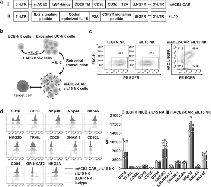
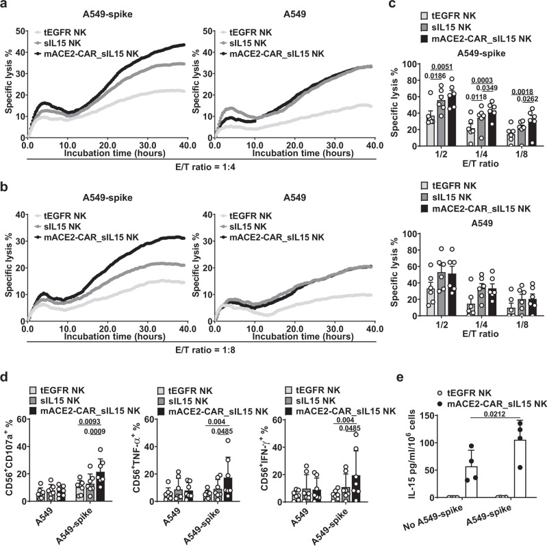
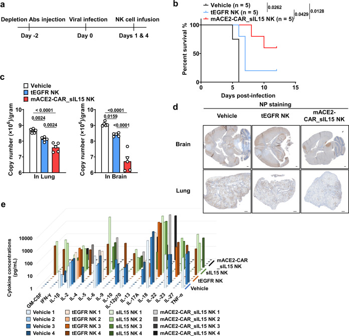
Engineered natural killer (NK) cells represent a promising option for immune therapy option due to their immediate availability in allogeneic settings. Severe acute diseases, such as COVID-19, require targeted and immediate intervention. Here we show engineering of NK cells to express (1) soluble interleukin-15 (sIL15) for enhancing their survival and (2) a chimeric antigen receptor (CAR) consisting of an extracellular domain of ACE2, targeting the spike protein of SARS-CoV-2. These CAR NK cells (mACE2-CAR_sIL15 NK cells) bind to VSV-SARS-CoV-2 chimeric viral particles as well as the recombinant SARS-CoV-2 spike protein subunit S1 leading to enhanced NK cell production of TNF-α and IFN-γ and increased in vitro and in vivo cytotoxicity against cells expressing the spike protein. Administration of mACE2-CAR_sIL15 NK cells maintains body weight, reduces viral load, and prolongs survival of transgenic mice expressing human ACE2 upon infection with live SARS-CoV-2. These experiments, and the capacity of mACE2-CAR_sIL15 NK cells to retain their activity following cryopreservation, demonstrate their potential as an allogeneic off-the-shelf therapy for COVID-19 patients who are faced with limited treatment options.


Conflict of interest statement

Drs. Caligiuri and Yu are co-founders of CytoImmune Therapeutics, Inc. The remaining authors have no conflicts of interest to disclose.

Figures

Fig. 1
Fig. 1. Generation of mACE2-CAR_sIL15 NK cells.
a Design of the mACE2-CAR and sIL15 retroviral vectors. The mACE2-CAR vector contains a mutant human ACE2, a human IgG1-hinge region, and the CD28 transmembrane region, followed by the intracellular domains of CD28 and CD3ζ linked to a T2A and a truncated (t) LNGFR (i). The sIL15 vector contains an IL-2 signaling peptide, a codon-optimized IL-15, and the GM-CSF receptor (CSF2R) signaling peptide, followed by a truncated (t) EGFR (ii). b Generation of mACE2-CAR_sIL15 NK cells. The pictures were created with BioRender.com. c The transduction efficiency of control tEGFR or sIL15 NK cells and mACE2-CAR_sIL15 NK cells was determined by flow cytometry. Control tEGFR or sIL15 NK cells expressed the tEGFR marker, while mACE2-CAR_sIL15 NK cells expressed the tLNGFR maker (representing mACE2-CAR expression) as well as the tEGFR marker (representing sIL15 expression). d The phenotypic analysis of mACE2-CAR_sIL15 NK cells and control tEGFR or sIL15 NK cells were determined by flow cytometry. Representative flow cytometry histograms and summary data from three different donors are shown. P values were determined by one-way ANOVA with multiple comparisons and adjusted by the Holm-Sidak method. Data are presented as mean ± SD. For mACE2-CAR_sIL15 NK cells, sIL15 expression is denoted as LNGFREGFR+, mACE2-CAR expression is denoted as LNGFR+EGFR, and sIL15 & mACE2-CAR_co-expression is denoted as LNGFR+EGFR+. Source data are provided as a Source Data file.
Fig. 2
Fig. 2. mACE2-CAR_sIL15 NK cells bind to SARS-CoV-2 spike protein and VSV-SARS-CoV-2 chimeric viral particles.
a Diagram of mACE2-CAR_sIL15 NK cells binding to spike S1-His-tagged recombinant protein. MFI = mean fluorescence intensity. The picture was created with BioRender.com. b, c Representative flow cytometry plots showing the results of mACE2-CAR_sIL15 NK cells binding to spike S1-His-tagged recombinant protein (b). LNGFR was used as a marker for mACE2-CAR-positive NK cells. Summary data from five different donors are shown in c. P values were determined by one-way ANOVA with multiple comparisons and adjusted by the Holm–Sidak method. Data are presented as mean ± SD. d Diagram of spike S1-His-tagged recombinant protein binding to VSV-SARS-CoV-2 chimeric viral particles, which are recognized by anti-spike and its corresponding fluor-conjugated secondary antibody. The picture was created with BioRender.com. e Representative flow cytometry plots showing the efficiency of control NK or mACE2-CAR_sIL15 NK cells binding to VSV-SARS-CoV-2 chimeric viral particles. f, g Representative flow cytometry plots showing the efficiency of CAR-negative or CAR-positive subsets in mACE2-CAR_sIL15 NK cells binding to VSV-SARS-CoV-2 chimeric viral particles. Summary data from five different donors are shown in g. P values were determined by one-way ANOVA with multiple comparisons and adjusted by the Holm-Sidak method. Data are presented as mean ± SD. Source data are provided as a Source Data file.
Fig. 3
Fig. 3. mACE2-CAR_sIL15 enhances NK cell cytotoxicity against target cells that express SARS-CoV-2 spike protein.
a Representative real-time cell analysis (RTCA) data showing NK cell cytotoxicity against A549-spike or parental A549 cells at an effector (E)/target (T) ratio of 1:4. b RTCA data showing NK cell cytotoxicity against A549-spike or parental A549 cells at an E/T ratio of 1:8. c Data from the RTCA assay analyzed at three different E/T ratios are summarized for cells from 6 donors. P values were determined with one-way ANOVA with multiple comparisons and adjusted by the Hochberg method. Data are presented as mean ± SEM. d Expression of CD107a, TNF-α, and IFN-γ was measured on transduced NK cells co-cultured with A549-spike or parental A549 cells for 4 h at an E/T ratio of 4:1. Data are summarized for NK cells from 7 donors. P values were determined by one-way ANOVA with multiple comparisons and adjusted by the Holm-Sidak or Dunn’s method. Data are presented as mean ± SD. e IL-15 production by ex vivo-expanded control tEGFR NK cells or mACE2-CAR_sIL15 NK cells cultured in the presence or absence of A549-spike cells for 72 h. Data are summarized for NK cells from four donors. P values were determined by one-way ANOVA with multiple comparisons and adjusted by the Holm–Sidak method. Data are presented as mean ± SD. Source data are provided as a Source Data file.
Fig. 4
Fig. 4. Freeze-thawed mACE2-CAR_sIL15 NK cells show effective anti-spike activity in vitro and in vivo.
a CAR expression in mACE2-CAR_sIL15 NK cells post-thaw, as determined by flow cytometry. Data are summarized for cells from three donors. P value was determined by two-tailed paired t test. Data are presented as mean ± SD. b Cell viability of mACE2-CAR_sIL15 NK cells as determined at the indicated time points post-thaw using the Muse Cell Analyzer. Data are summarized for NK cells from three donors. P values were determined by two-way ANOVA with repeated measures and adjusted by the Holm-Sidak method. Data are presented as mean ± SD. c Lysis of freeze-thawed control NK or mACE2-CAR_sIL15 NK cells that were co-cultured with A549-spike or parental A549 cells and analyzed with real-time cell analysis (RTCA). Data are summarized for NK cells from three donors. P values were determined with a two-tailed paired t test. Data are presented as mean ± SEM. d Scheme for in vivo studies using NSG mice. e Tumor growth in NSG mice inoculated with firefly luciferase-labeled A549-spike cells, as monitored by changes in tumor bioluminescence. Colors indicate intensity of luminescence. f Summary of tumor burden data from e (four mice/group). P values were determined by one-way ANOVA with with multiple comparisons and adjusted by the Holm-Sidak method on day 19. Data are presented as mean ± SEM. g After hematoxylin and eosin (H&E) staining of lung tissues, tumor burden was identified and evaluated as the number of tumor nodules, which is defined as ≥50 µm. Four fields of Regions of Interest (ROIs) per lung were selected, and the number of lung tumor nodules per four fields was counted. Summary data are shown (four mice/group). P values were determined by one-way ANOVA with multiple comparisons and adjusted by the Holm-Sidak method. Data are presented as mean ± SD. Source data are provided as a Source Data file.
Fig. 5
Fig. 5. mACE2-CAR_sIL15 NK cells protect against live SARS-CoV-2 infection in the K18-hACE2 humanized mouse model.
a Scheme for in vivo studies using K18-hACE2 humanized transgenic mice. b K18-hACE2 humanized transgenic mice were infected with 1 × 103 plaque-forming units (PFU) of SARS-CoV-2 prior to being treated with the vehicle (PBS), control tEGFR NK cells, or mACE2-CAR_sIL15 NK cells. Survival of mice is summarized. N = 5 mice/group. P values were determined by Kaplan–Meier survival analysis and calculated by the Gehan-Breslow-Wilcoxon test (two-sided). c Viral RNA levels are shown for brain and lung tissues of mice infected i.n. with 1 × 103 plaque-forming units (PFU) of SARS-CoV-2. N = 5 mice/group. P values were determined by one-way ANOVA with multiple comparisons and adjusted by the Holm-Sidak method. Data are presented as mean ± SEM. d Representative images of the three groups in b show immunohistochemistry (IHC) staining of coronavirus nucleocapsid protein (NP). Scale bars, 400 μm. e Release assay of various cytokines into plasma of K18-hACE2 transgenic mice. Mice were infected with 1 × 103 PFU of live SARS-CoV-2 prior to being treated with the vehicle (PBS), control tEGFR NK cells, control sIL15 NK cells, or mACE2-CAR_sIL15 NK cells. Four days later, all mice were sacrificed to collect blood plasma to measure levels of the indicated cytokines by a cytokine release Luminex assay. N = 4 mice/group. Data are presented as an average of two replicates for each mouse. Source data are provided as a Source Data file.

References

    1. Zhu N, et al. A novel coronavirus from patients with pneumonia in China, 2019. N. Engl. J. Med. 2020;382:727–733. doi: 10.1056/NEJMoa2001017. - DOI - PMC - PubMed
    1. Organization WHO. Coronovirus disease (COVID-19) pandemic. (2022).
    1. Spinner CD, et al. Effect of remdesivir vs standard care on clinical status at 11 days in patients with moderate COVID-19: a randomized clinical trial. JAMA. 2020;324:1048–1057. doi: 10.1001/jama.2020.16349. - DOI - PMC - PubMed
    1. Wang Y, et al. Remdesivir in adults with severe COVID-19: a randomised, double-blind, placebo-controlled, multicentre trial. Lancet. 2020;395:1569–1578. doi: 10.1016/S0140-6736(20)31022-9. - DOI - PMC - PubMed
    1. Group R. C. et al. Dexamethasone in hospitalized patients with Covid-19 - preliminary report. N. Engl. J. Med. 384, 693–704 (2020). - PMC - PubMed

Publication types

Substances