. 2022 May 11;12(1):7746.
doi: 10.1038/s41598-022-11967-2.

Rehearsal simulation to determine the size of device for left atrial appendage occlusion using patient-specific 3D-printed phantoms

Affiliations

Rehearsal simulation to determine the size of device for left atrial appendage occlusion using patient-specific 3D-printed phantoms

Dayeong Hong et al. Sci Rep. .

Abstract

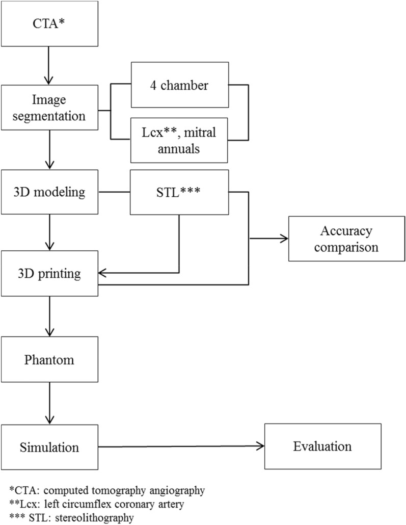
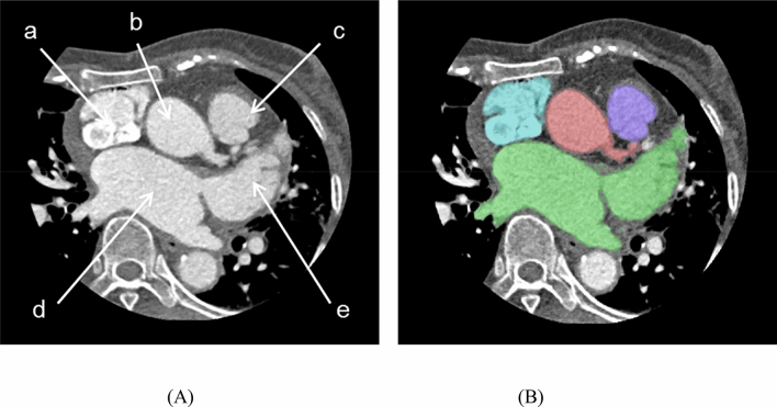
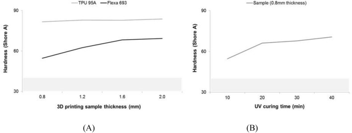
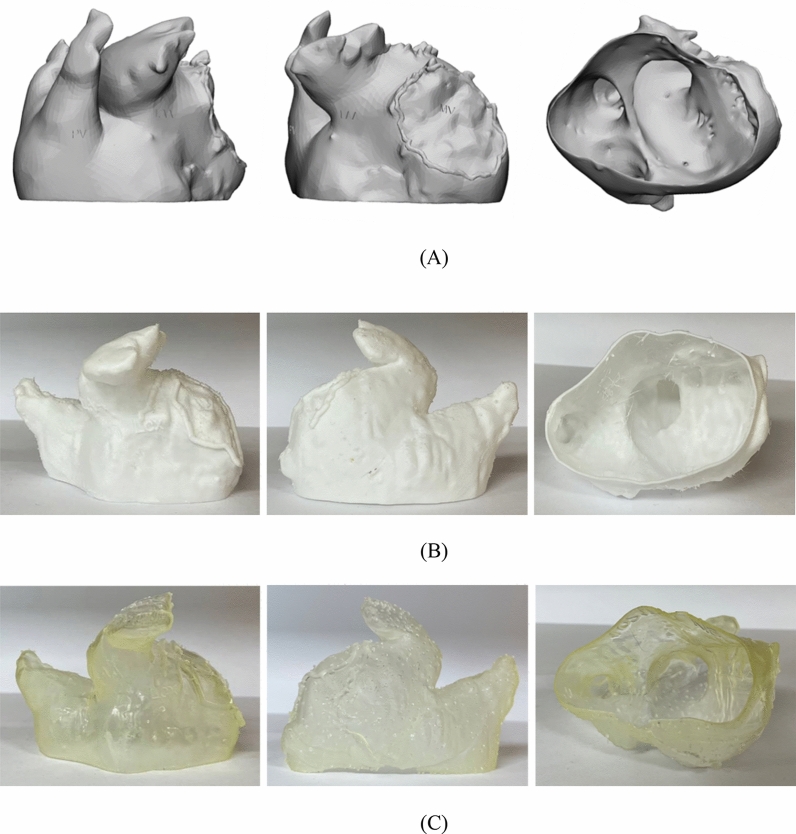
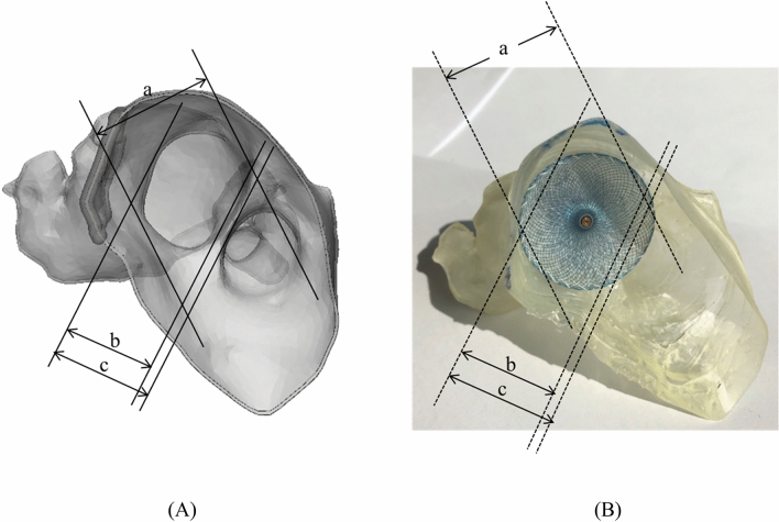
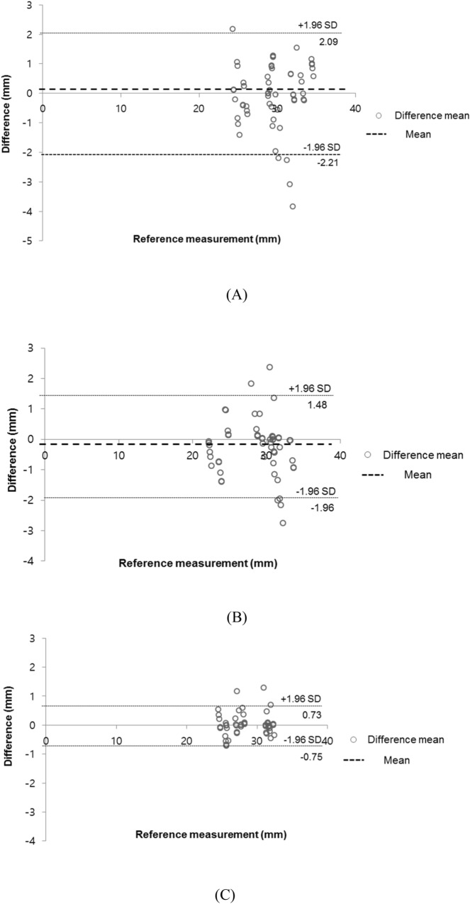
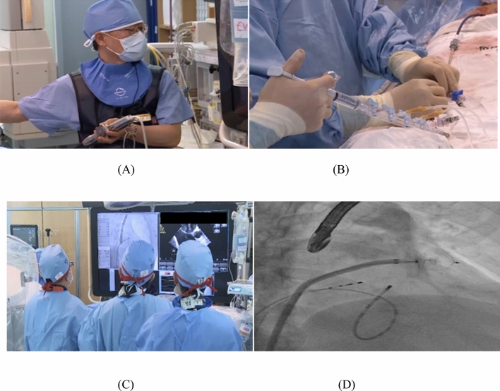
Left atrial appendage (LAA) occlusion (LAAO) is used to close the finger-like extension from the left atrium with occlusion devices to block the source of thrombosis. However, selection of the devices size is not easy due to various anatomical changes. The purpose of this study is patient-specific, computed tomography angiography (CTA)-based, three-dimensionally (3D) printed LAAO phantoms were applied pre-procedure to determine the size. Ten patients were enrolled prospectively in March 2019 and December 2020. The cardiac structure appearing in CTA was first segmented, and the left atrium and related structures in the LAAO procedure were modeled. The phantoms were fabricated using two methods of fused deposition modeling (FDM) and stereolithography (SLA) 3D printers with thermoplastic polyurethane (TPU) and flexible resin materials and evaluated by comparing their physical and material properties. The 3D-printed phantoms were directly used to confirm the shape of LAA, and to predict the device size for LAAO. In summary, the shore A hardness of TPU of FDM was about 80-85 shore A, and that of flexible resin of SLA was about 50-70 shore A. The measurement error between the STL model and 3D printing phantoms were 0.45 ± 0.37 mm (Bland-Altman, limits of agreement from - 1.8 to 1.6 mm). At the rehearsal, the estimations of device sizes were the exact same with those in the actual procedures of all 10 patients. In conclusion, simulation with a 3D-printed left atrium phantom could be used to predict the LAAO insertion device size accurately before the procedure.


Conflict of interest statement

The authors declare no competing interests.

Figures

Figure 1
Figure 1
Overall workflow for developing rehearsal phantom for LAAO with 3D printing and medical images. 3D three-dimensional, CTA computed tomography angiography, LAAO left atrial appendage occlusion, Lcx left circumflex coronary artery, STL stereolithography.
Figure 2
Figure 2
Segmentation based on cardiac anatomy by cardiac CT angiography, including: (A) CT image based cardiac anatomy: (a) cavoatrial junction, (b) ascending aorta, (c) main pulmonary artery, (d) left atrium, (e) left atrial appendage; and (B) Segmentation based on CT angiography. CT computed tomography (Spectral 3, Filter B, Philips, Best, The Netherlands).
Figure 3
Figure 3
Comparison of mechanical properties of 3D printing materials. The gray zone in both graphs is human cardiac tissue to shore A hardness (mean) of about 40. (A) Hardness with different thickness values of two FDM printing materials. (B) Hardness according to UV curing time of one material. 3D three-dimensional, FDM fused deposition modeling, UV ultraviolet.
Figure 4
Figure 4
LAAO phantom made with two types of 3D printers. (A) 3D modeling (B) FDM, (C) SLA. 3D three-dimensional, FDM fused deposition modeling, LAAO left atrial appendage occlusion, SLA stereolithography.
Figure 5
Figure 5
Measurements of shape accuracy between 3D model and 3D-printed phantom. (A) The 3D model (STL) with three landmarks specified for evaluating measurement error. (B) The 3D-printed phantom with three landmarks specified for evaluating measurement error. (a, diameter of the horizontal zone; b, LAA ostium; c, vertical zone). 3D three-dimensional, LAA left atrial appendage, STL stereolithography.
Figure 6
Figure 6
The Bland–Altman analysis to evaluate differences between the 3D model (STL) and the 3D-printed phantom. (A) Diameter of the horizontal zone, (B) LAA ostium, (C) Vertical zone. (SPSS version 25.00; IBM Corp., Armonk, NY, USA). 3D three-dimensional, STL Stereolithography, LAA left atrial appendage.
Figure 7
Figure 7
LAAO was performed using a device of the predicted size. (A) A cardiologist performing TEE during the procedure. (B) Another cardiologist injecting saline, medicine, and medium contrast through the manifold. (C) Checking of fluoroscopy and TEE in real time during the procedure. (D) Confirmation of the device insertion predicted by fluoroscopy correctly into the LAA. LAA left atrial appendage, LAAO left atrial appendage occlusion, TEE transesophageal echocardiography.

Similar articles

Cited by

References

    1. Whitlock, R. P. Stroke and Left atrial appendage occlusion in cardiac surgery (2013).
    1. Burysz M, Litwinowicz R, Burysz A, Ogorzeja W, Bartuś K. Causes of death and morbidity in patients with atrial fibrillation after left atrial appendage occlusion. Kardiol. Pol. 2019;77:1047–1054. doi: 10.33963/KP.14966. - DOI - PubMed
    1. Horstmann S, et al. Left atrial appendage occlusion in atrial fibrillation after intracranial hemorrhage. Neurology. 2014;82:135–138. doi: 10.1212/WNL.0000000000000022. - DOI - PMC - PubMed
    1. Briceno, D. F. et al. Left atrial appendage occlusion device and novel oral anticoagulants versus warfarin for stroke prevention in nonvalvular atrial fibrillation: systematic review and meta-analysis of randomized controlled trials. Circ. Arrhythmia Electrophysiol.8, 1057–1064 (2015). - PubMed
    1. Bayard YL, et al. PLAATO (Percutaneous Left Atrial Appendage Transcatheter Occlusion) for prevention of cardioembolic stroke in non-anticoagulation eligible atrial fibrillation patients: Results from the European PLAATO study. EuroIntervention. 2010;6:220–226. doi: 10.4244/EIJV6I2A35. - DOI - PubMed

Publication types

MeSH terms