. 2022 Dec;71(12):2969-2983.
doi: 10.1007/s00262-022-03209-1. Epub 2022 May 12.

In vitro and in vivo evaluation of DC-targeting PLGA nanoparticles encapsulating heparanase CD4+ and CD8+ T-cell epitopes for cancer immunotherapy

Affiliations

In vitro and in vivo evaluation of DC-targeting PLGA nanoparticles encapsulating heparanase CD4+ and CD8+ T-cell epitopes for cancer immunotherapy

Xu-Dong Tang et al. Cancer Immunol Immunother. 2022 Dec.

Abstract

Heparanase has been identified as a universal tumor-associated antigen, but heparanase epitope peptides are difficult to recognize. Therefore, it is necessary to explore novel strategies to ensure efficient delivery to antigen-presenting cells. Here, we established a novel immunotherapy model targeting antigens to dendritic cell (DC) receptors using a combination of heparanase CD4+ and CD8+ T-cell epitope peptides to achieve an efficient cytotoxic T-cell response, which was associated with strong activation of DCs. First, pegylated poly(lactic-coglycolic acid) (PLGA) nanoparticles (NPs) were used to encapsulate a combined heparanase CD4+ and CD8+ T-cell epitope alone or in combination with Toll-like receptor 3 and 7 ligands as a model antigen to enhance immunogenicity. The ligands were then targeted to DC cell-surface molecules using a DEC-205 antibody. The binding and internalization of these PLGA NPs and the activation of DCs, the T-cell response and the tumor-killing effect were assessed. The results showed that PLGA NPs encapsulating epitope peptides (mHpa399 + mHpa519) could be targeted to and internalized by DCs more efficiently, stimulating higher levels of IL-12 production, T-cell proliferation and IFN-γ production by T cells in vitro. Moreover, vaccination with DEC-205-targeted PLGA NPs encapsulating combined epitope peptides exhibited higher tumor-killing efficacy both in vitro and in vivo. In conclusion, delivery of PLGA NP vaccines targeting DEC-205 based on heparanase CD4+ and CD8+ T-cell epitopes are suitable immunogens for antitumor immunotherapy and have promising potential for clinical applications.

Keywords: Dendritic cells; Heparanase; Nanoparticles; Targeting; Tumor immunotherapy.


Conflict of interest statement

The authors declare that there are no conflict of interest.

Figures

Fig. 1
Fig. 1
Schematic diagram of PLGA NP encapsulating heparanase epitope peptides targeting DEC-205 on mouse DCs. A PLGA NP vaccines were generated in combination with different heparanase epitope peptides as indicated. B Image analysis revealed the morphology of NP (scale bar, 0.5 μm; magnification, 25,000×)
Fig. 2
Fig. 2
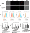
Uptake efficacy of NPs. A BMDCs were incubated with Lightning-Link® Rapid DyLight® 680 labeled NP-(mHpa519)-αDEC-205 or NP-(mHpa519)-isotype (20 μg/mL) at 37 °C for 3 h, and then stained with FITC-anti-mouse CD11c and DAPI. Images were captured with a Leica scanning microscope (Leica TCS-SP5). WL panel (white light), DAPI panel (BMDCs nuclear stained with DAPI); CD11c panel (FITC-anti-mouse CD11c fluorescent images); NP panel (Lightning-Link® Rapid DyLight® 680 labeled NP fluorescent images); merge panel, merged images. B The uptake efficacy was evaluated by histograms. BMDCs were incubated for 3 h at either 4 °C (binding analysis) or 37 °C (internalization analysis) with 20 μg/mL NPs. Then washed to remove unbound NPs. For internalization assay, the BMDCs were washed by acidic PBS (pH = 2) to remove the bound but not internalized NP. The fluorescence intensity was measured by a BD FACsAria cytometer. (C and D) BMDCs were incubated for 3 h at either 4 °C (binding analysis) or 37 °C (internalization analysis) with 1, 5, 10, 20 μg/mL NPs. Then washed to remove unbound NPs. For internalization assay, the BMDCs were washed by acidic PBS (pH = 2) to remove the bound but not internalized NPs. The mean fluorescence intensity (MFI) was measured by a BD FACsAria cytometer. (E) The internalization ratio was calculated following the formula: %internalization = [(MFI in 37 °C-background)]/[(MFI on ice)-background] × 100%. Data are representative of three independent experiments. *p < 0.05; **p < 0.01 (by student’s t test)
Fig. 3
Fig. 3
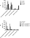
Activation of DCs in vitro by targeted PLGA NP. C57BL/6 BMDCs (100,000 cells/well) were incubated with increasing concentration of NP-(mHpa519 + 399)-αDEC-205, or NP-(mHpa519)-αDEC-205, or NP-(NC)- αDEC-205, NP-(mHpa519)-isotype, for 24 h at 37 °C. Culture supernatants were harvested, and the amount of IL-12 was determined by ELISA (A). Differences in cytokine production were analyzed applying two-way ANOVA analysis. Data shown are mean ± SD from one representative experiment out of 3 independent experiments. B Dendritic cells were pre-incubated for 1 h at 37 °C with titrated amounts of Cytochalasin D followed by a 24 h incubation with 10 μg/ml of NP-(mHpa399 + 519)-αDEC-205, NP-(mHpa519)-αDEC-205, or non-targeted. Indicated amounts of Cytochalasin D were maintained during the 24 h incubation with NP. After incubation, culture supernatants were harvested and analyzed for IL-12 amounts by ELISA
Fig. 4
Fig. 4
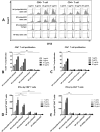
DEC-205 targeted NP containing heparanase epitope peptides, poly I:C, and R848 improved T cell stimulatory capacity. A BMDCs were incubated for 3 h at 37 °C with 20 μg/mL NP-(mHpa519 + 399)-αDEC-205, or NP-(mHpa519)-αDEC-205, or NP-(mHpa519)-isotype, or NP-(NC)-αDEC-205. Splenocytes were labeled at 107 cells/mL with 5 μM CFSE and then incubated with NPs loaded BMDCs for 6 days at (1 BMDC: 5 splenocytes). Thereafter, T cells were stained with APC-anti-mouse CD3, PerCP-anti-mouse CD4 and PE-anti-mouse CD8. T cell proliferation was measured by a BD FACsAria cytometer. B, C NP-(mHpa519 + 399)-αDEC-205, or NP-(mHpa519)-αDEC-205 was taken up by BMDCs and mediated activation of heparanase-specific T cells. Data shown represent the mean ± SD from triplicates of characteristic of three independent experiments (*p < 0.05, **p < 0.01). (D and E) CD8+ T cells or CD4+ T cells were sorted separately by flow cytometry. IFN-γ secreted by CD8+ T cells or CD4+ T cells was evaluated. Data shown represent the mean ± SD from triplicates of characteristic of three independent experiments (*p < 0.05, **p < 0.01)
Fig. 5
Fig. 5
The killing effect of CTLs induced by DEC-205 targeted NP containing heparanase epitope. C57BL/6 mice were immunized thrice at 7-day intervals by s.c. injection of 5 × 105 NP-(mHpa399 + 519)-αDEC-205, or PLGA-(mHpa519)-αDEC-205, or non-targeted NP-pulsed dendritic cells. On day 28, mice splenocytes were served as effectors. Standard 51Cr-release assays were performed to test for their cytotoxic activity against B16 melanoma cells (A) and Lewis lung cancer cells (B) at various effector: target (E/T) ratios. Data shown are mean ± SEM of three independent experiments (**p < 0.01). C, D Mouse primary aortic endothelial cells (PAEC) were transfected with heparanase expression plasmid and control vectors. Standard 51Cr-release assays were performed as above
Fig. 6
Fig. 6
DEC-205 targeted NP containing heparanase epitope peptides vaccination reduce tumor burden. Mice were vaccinated by NP-(mHpa519 + 399)-αDEC-205, or NP-(mHpa519)-αDEC-205, or NP-(NC)- αDEC-205, or NP-(mHpa519)-isotype loading BMDCs 3 times at 7-day intervals. Seven days after the last vaccination, mice were challenged with 1 × 106 B16 melanoma cells by inoculated s.c. in the left flank. Tumor growth (A, B) and animal survival (C) were monitored. Mice were immunized as (A), and then mice were challenged with 1 × 106 Lewis cancer cells by i.v. injection through tail vein. Lewis cancer pulmonary metastasis (D, E) and animal survival (F) were monitored. Differences in tumor sizes and metastasis nodules per group were analyzed by regular two-way ANOVA with Bonferroni posttests to calculate the difference in mean values at each time point. Animal survival per group was assessed using log-rank (Mantel–Cox) test, **p < 0.001, *p < 0.05

Similar articles

Cited by

References

    1. Schreiber RD, Old LJ, Smyth MJ. Cancer immunoediting: integrating immunity's roles in cancer suppression and promotion. Science. 2011;331(6024):1565–1570. doi: 10.1126/science.1203486. - DOI - PubMed
    1. Vesely MD, Kershaw MH, Schreiber RD, Smyth MJ. Natural innate and adaptive immunity to cancer. Annu Rev Immunol. 2011;29:235–271. doi: 10.1146/annurev-immunol-031210-101324. - DOI - PubMed
    1. Finn OJ. Vaccines for cancer prevention: a practical and feasible approach to the cancer epidemic. Cancer Immunol Res. 2014;2(8):708–713. doi: 10.1158/2326-6066.CIR-14-0110. - DOI - PMC - PubMed
    1. Coulie PG, Van den Eynde BJ, van der Bruggen P, Boon T. Tumour antigens recognized by T lymphocytes: at the core of cancer immunotherapy. Nat Rev Cancer. 2014;14(2):135–146. doi: 10.1038/nrc3670. - DOI - PubMed
    1. Hinrichs CS, Restifo NP. Reassessing target antigens for adoptive T-cell therapy. Reassessing target antigens for adoptive T-cell therapy. Nat Biotechnol. 2013;31(11):999–1008. doi: 10.1038/nbt.2725. - DOI - PMC - PubMed