

Video: Clinical evaluation of a laparoscopic hyperspectral imaging system
- PMID: 35546207
- PMCID: PMC9485189
- DOI: 10.1007/s00464-022-09282-y
Video: Clinical evaluation of a laparoscopic hyperspectral imaging system
Abstract
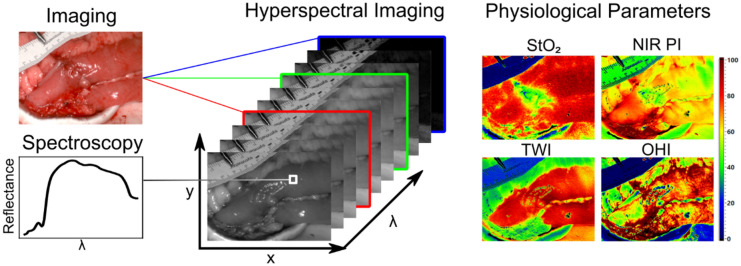
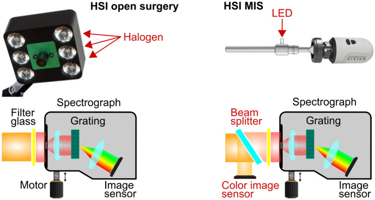
Background: Hyperspectral imaging (HSI) during surgical procedures is a new method for perfusion quantification and tissue discrimination. Its use has been limited to open surgery due to large camera sizes, missing color video, or long acquisition times. A hand-held, laparoscopic hyperspectral camera has been developed now to overcome those disadvantages and evaluated clinically for the first time.
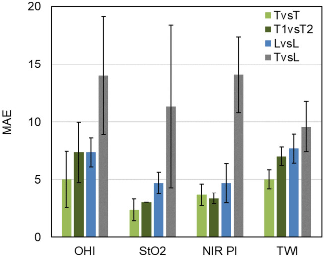
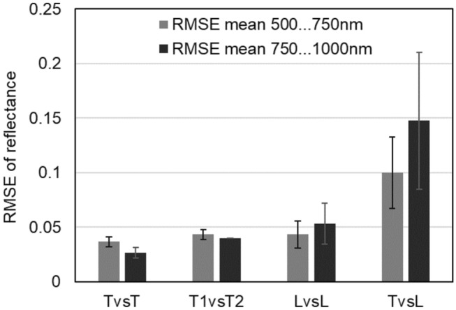
Methods: In a clinical evaluation study, gastrointestinal resectates of ten cancer patients were investigated using the laparoscopic hyperspectral camera. Reference data from corresponding anatomical regions were acquired with a clinically approved HSI system. An image registration process was executed that allowed for pixel-wise comparisons of spectral data and parameter images (StO2: oxygen saturation of tissue, NIR PI: near-infrared perfusion index, OHI: organ hemoglobin index, TWI: tissue water index) provided by both camera systems. The mean absolute error (MAE) and root mean square error (RMSE) served for the quantitative evaluations. Spearman's rank correlation between factors related to the study design like the time of spectral white balancing and MAE, respectively RMSE, was calculated.
Results: The obtained mean MAEs between the TIVITA® Tissue and the laparoscopic hyperspectral system resulted in StO2: 11% ± 7%, NIR PI: 14±3, OHI: 14± 5, and TWI: 10 ± 2. The mean RMSE between both systems was 0.1±0.03 from 500 to 750 nm and 0.15 ±0.06 from 750 to 1000 nm. Spearman's rank correlation coefficients showed no significant correlation between MAE or RMSE and influencing factors related to the study design.
Conclusion: Qualitatively, parameter images of the laparoscopic system corresponded to those of the system for open surgery. Quantitative deviations were attributed to technical differences rather than the study design. Limitations of the presented study are addressed in current large-scale in vivo trials.
Keywords: Clinical evaluation study; Gastrointestinal surgery; Hyperspectral imaging; Laparoscopic surgery; Minimally invasive surgery.
© 2022. The Author(s).
Conflict of interest statement
Annekatrin Pfahl, Hannes Köhler, Madeleine T. Thomaßen, Marianne Maktabi, Albrecht M. Bloße, Matthias Mehdorn, Orestis Lyros, Yusef Moulla, Stefan Niebisch, Boris Jansen-Winkeln, Claire Chalopin, and Ines Gockel have no conflict of interest or financial ties to disclose. KARL STORZ SE & Co. KG and Diaspective Vision GmbH provided the equipment for the measurements
Figures
References
-
- Jansen-Winkeln B, Germann I, Köhler H, Mehdorn M, Maktabi M, Sucher R, Barberio M, Chalopin C, Diana M, Moulla Y, Gockel I. Comparison of hyperspectral imaging and fluorescence angiography for the determination of the transection margin in colorectal resections—a comparative study. Int J Colorectal Dis. 2020 doi: 10.1007/s00384-020-03755-z. - DOI - PMC - PubMed
-
- Barberio M, Felli E, Seyller E, Longo F, Chand M, Gockel I, Geny B, Swanström L, Marescaux J, Agnus V, Diana M. Quantitative fluorescence angiography versus hyperspectral imaging to assess bowel ischemia: a comparative study in enhanced reality. Surgery. 2020;168:178–184. doi: 10.1016/j.surg.2020.02.008. - DOI - PubMed
Publication types
MeSH terms
Substances
LinkOut - more resources
Full Text Sources
Research Materials
Miscellaneous
