. 2023 Jan-Feb;25(1):103-112.
doi: 10.4103/aja202226.

Epididymis cell atlas in a patient with a sex development disorder and a novel NR5A1 gene mutation

Affiliations

Epididymis cell atlas in a patient with a sex development disorder and a novel NR5A1 gene mutation

Jian-Wu Shi et al. Asian J Androl. 2023 Jan-Feb.

Abstract

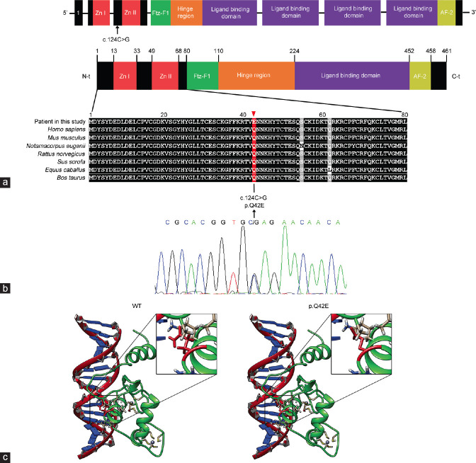
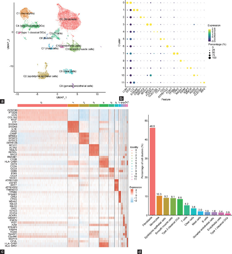
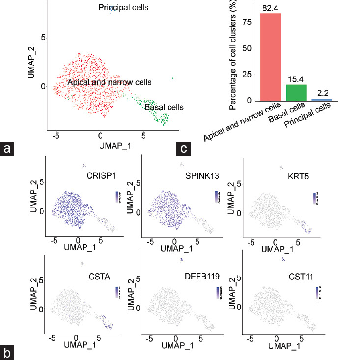
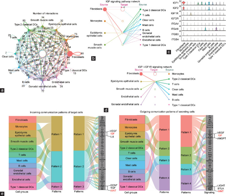
This study aims to characterize the cell atlas of the epididymis derived from a 46,XY disorders of sex development (DSD) patient with a novel heterozygous mutation of the nuclear receptor subfamily 5 group A member 1 (NR5A1) gene. Next-generation sequencing found a heterozygous c.124C>G mutation in NR5A1 that resulted in a p.Q42E missense mutation in the conserved DNA-binding domain of NR5A1. The patient demonstrated feminization of external genitalia and Tanner stage 1 breast development. The surgical procedure revealed a morphologically normal epididymis and vas deferens but a dysplastic testis. Microfluidic-based single-cell RNA sequencing (scRNA-seq) analysis found that the fibroblast cells were significantly increased (approximately 46.5%), whereas the number of main epididymal epithelial cells (approximately 9.2%), such as principal cells and basal cells, was dramatically decreased. Bioinformatics analysis of cell-cell communications and gene regulatory networks at the single-cell level inferred that epididymal epithelial cell loss and fibroblast occupation are associated with the epithelial-to-mesenchymal transition (EMT) process. The present study provides a cell atlas of the epididymis of a patient with 46,XY DSD and serves as an important resource for understanding the pathophysiology of DSD.

Keywords: NR5A1; disorders of sex development; human epididymis; scRNA-seq.


Conflict of interest statement

None

Figures

Figure 1
Figure 1
Schematic diagram of the novel mutation site of NR5A1 gene. (a) Schematic graph for the NR5A1 showing the location of the mutation. (b) The mutated p.Q42E was located between two zinc domains, which is important for DNA-binding activity. The alignment of NR5A1 of p.Q42E and its neighboring AAs are displayed in the middle panel. The sequence of the affected AA is conserved. Sanger sequencing showing the c.124C>G substitution is depicted below. (c) Predicted structures of the protein encoded by NR5A1 of normal and mutation p.Q42E. NR5A1: nuclear receptor subfamily 5 group A member 1; AA: amino acid; N-t: NH2-terminal; C-t: carboxyl-terminal; AF-2: activation function-2 domain; Ftz-F1: Fushi Tarazu factor 1.
Figure 2
Figure 2
Overview of scRNA sequencing for the human epididymis with the NR5A1 gene p.Q42E mutation. (a) UMAP visualization of the twelve cell clusters annotated in the patients’ epididymis after erythrocyte filtration. (b) Cell annotation of each epididymal cell population based on cell-type-specific marker genes from the literature (expression values are centered and scaled by row). (c) The top 5 marker genes of the heatmap showed for each population (expression scaled by row). (d) The cell proportion of each cluster was displayed. NR5A1: nuclear receptor subfamily 5 group A member 1; UMAP: uniform manifold approximation and projection; scRNA: single cell RNA.
Figure 3
Figure 3
Features of epididymis epithelial cell subpopulations. (a) UMAP graph of the subclusters in the epididymal epithelial cells. Three subclusters of epididymis epithelial cells were identified. (b) The proportion of each epithelial cell subpopulation is illustrated. (c) Representative cell-specific marker illustrated by UMAP of epididymal epithelial cell clusters (color values are expression). UMAP: uniform manifold approximation and projection; CRISP1: cysteine-rich secretory protein 1; SPINK13: serine peptidase inhibitor Kazal type 13; KRT5: keratin 5; CSTA: cystatin A; DEFB119: defensin beta 119; CST11: cystatin 11.
Figure 4
Figure 4
CellChat analysis between fibroblasts and epididymis epithelial cells. (a) Overview of pronounced ligand–receptor pairs in cell clusters. The thickness of the line represents ligand–receptor pair number. (b) Hierarchical plot representing the deductive cell communications for the IGF signaling pathway. (c) Violin plot displaying the expression of IGF1 signaling genes in each cell cluster. (d) Hierarchical plot showing the interactions for IGF1-IGF1R signaling. (e) Alluvial plot showing the incoming and outgoing signaling patterns of cell groups. The inferred outgoing communication patterns of secreting cells and incoming patterns of target cells suggest the correlation between the inferred potential cell groups, patterns, and signaling pathways. IGF1R: insulin-like growth factor 1 receptor; DC: dendritic cell; IGF: insulin-like growth factor; ITGB: integrin subunit beta; VEGF: vascular endothelial growth factor; ANGPT: angiopoietin; GDNF: glial cell-derived neurotrophic factor; NGF: nerve growth factor; WNT: Wingless; BTLA: B and T lymphocyte associated; LIGHT: TNF superfamily member 14.
Figure 5
Figure 5
Transcription factor regulatory networks derived from SCENIC analysis. (a) Regulon activity for the epididymal cell clusters. The color bar indicates cell clusters. The on/off state of the cluster-specific regulons are displayed by heatmap. Black represents the state in which the regulon was activated, and white represents the state in which the regulon was inactivated. The HOX gene family is magnified. (b) Heatmap showing the expression level of the HOX gene family in each population. (c) Cytoscape visualization revealed the networks of transcriptional factors and target genes of the HOX gene family. Biological processes and motif sequences of target genes for HOX transcription factors were listed. g: targeting gene; HOX: homeobox; MF: molecular function; SCENIC: Single-Cell rEgulatory Network Inference and Clustering.

Similar articles

Cited by

References

    1. Hughes IA, Houk C, Ahmed SF, Lee PA Lawson Wilkins Pediatric Endocrine Society/European Society for Paediatric Endocrinology Consensus Group. Consensus statement on management of intersex disorders. J Pediatr Urol. 2006;2:148–62. - PubMed
    1. Nabhan ZM, Lee PA. Disorders of sex development. Curr Opin Obstet Gynecol. 2007;19:440–5. - PubMed
    1. Aydin BK, Saka N, Bas F, Bas EK, Coban A, et al. Frequency of ambiguous genitalia in 14,177 newborns in Turkey. J Endocr Soc. 2019;3:1185–95. - PMC - PubMed
    1. Ohnesorg T, Vilain E, Sinclair AH. The genetics of disorders of sex development in humans. Sex Dev. 2014;8:262–72. - PubMed
    1. Pasterski V, Prentice P, Hughes IA. Impact of the consensus statement and the new DSD classification system. Best Pract Res Clin Endocrinol Metab. 2010;24:187–95. - PubMed

Substances