

Role of Vitamin D as Protective Agent against Induced Liver Damage in Male Rats
- PMID: 35546993
- PMCID: PMC9083877
- DOI: 10.22092/ari.2021.356357.1824
Role of Vitamin D as Protective Agent against Induced Liver Damage in Male Rats
Abstract
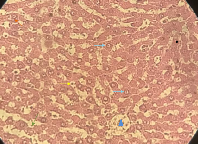
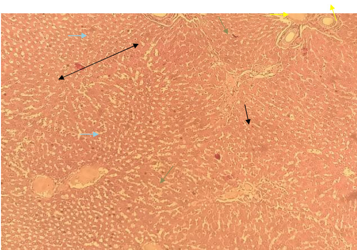
One of the main factors which played a key role in the prevention of liver disorders such as hepatic inflammation, fibrosis, and carcinogenesis would be the vitamin D axis. Therefore, the current research was designed to evaluate the role of Vitamin D (Vit D) as a protective agent against liver damage caused by Thioacetamide (TAA). In the current study, 18 male Wistar rats were randomly allocated into three equal groups (n=6): in group 1(G1) the animals were considered as the control group and did not receive any supplement in drinking water; in group 2 (G2) TAA was administrated to the drinking water at a dose of 300 mg/L; in group 3 (G3) TAA was administrated to the drinking water at a dose of 300 mg/L plus vitamin D at a dose of 0.5 mg/100g body (intraperitoneal) for 8 weeks. At the end of the experiment, the animals were sacrificed and the liver was dissected and removed for histopathology. Histopathological evaluations were used to evaluate the possible adverse effects of TAA on the liver. Several hepatic damages were observed in the G2 group such as lobular disorder, some degrees of degeneration in hepatocytes and enlargement of the hepatic capillaries, and focal necrotic areas. Hepatic fibrosis was observed around portal areas and central veins. Bridging fibrous septa were formed between portal veins. The recorded data in this study showed that Vit D has some beneficial effects in protecting the liver from fibrosis and toxic damages. The recorded data showed that liver damages in the G3 group were partially prevented or cured. In conclusion, it is evident that the Vit D played a pivotal role as an antioxidant and anti-fibrotic agent, therefore it would be the best supplement for liver protection against damages due to toxin entrance into the animal's body.
Keywords: Histopathological examinations; Liver fibrosis; Thioacetamide; Vitamin D.
Figures


References
-
- Ichimura R, Mizukami S, Takahashi M, Taniai E, Kemmochi S, Mitsumori K, et al. Disruption of Smad-dependent signaling for growth of GST-P-positive lesions from the early stage in a rat two-stage hepatocarcinogenesis model. Toxicol Appl Pharmacol. 2010;246(3):128–40. - PubMed
-
- JI PB, MM AH. Experimental thioacetamide-induced cirrhosis of the liver. Histol Histopathol. 1991;6(1):95–100. - PubMed
-
- Chilakapati J, Shankar K, Korrapati MC, Hill RA, Mehendale HM. Saturation toxicokinetics of thioacetamide: role in initiation of liver injury. Drug Metab Dispos. 2005;33(12):1877–85. - PubMed
-
- Natarajan SK, Thomas S, Ramamoorthy P, Basivireddy J, Pulimood AB, Ramachandran A, et al. Oxidative stress in the development of liver cirrhosis: a comparison of two different experimental models. J Gastroenterol Hepatol. 2006;21(6):947–57. - PubMed
MeSH terms
Substances
LinkOut - more resources
Full Text Sources
Medical
