

Personalized in vitro Extracellular Matrix Models of Collagen VI-Related Muscular Dystrophies
- PMID: 35547158
- PMCID: PMC9081367
- DOI: 10.3389/fbioe.2022.851825
Personalized in vitro Extracellular Matrix Models of Collagen VI-Related Muscular Dystrophies
Abstract
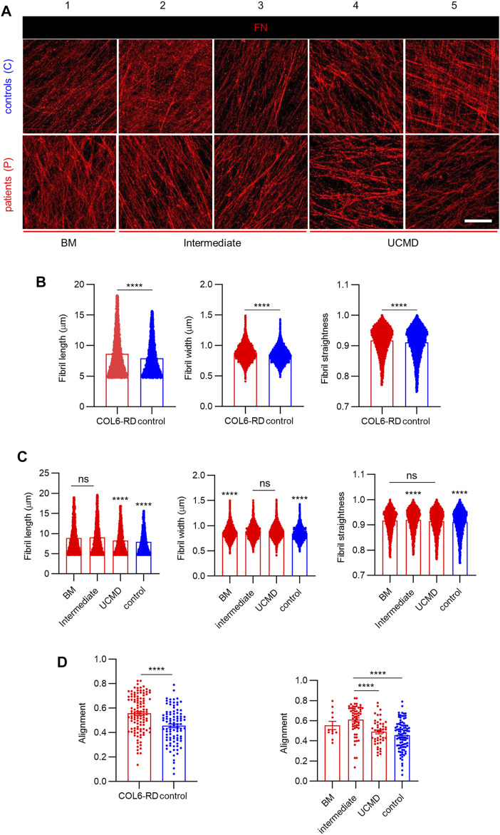
Collagen VI-related dystrophies (COL6-RDs) are a group of rare congenital neuromuscular dystrophies that represent a continuum of overlapping clinical phenotypes that go from the milder Bethlem myopathy (BM) to the severe Ullrich congenital muscular dystrophy, for which there is no effective treatment. Mutations in one of the three Collagen VI genes alter the incorporation of this protein into the extracellular matrix (ECM), affecting the assembly and the structural integrity of the whole fibrillar network. Clinical hallmarks of COL6-RDs are secondary to the ECM disruption and include muscle weakness, proximal joint contractures, and distal hyperlaxity. Although some traits have been identified in patients' ECMs, a correlation between the ECM features and the clinical phenotype has not been established, mainly due to the lack of predictive and reliable models of the pathology. Herein, we engineered a new personalized pre-clinical model of COL6-RDs using cell-derived matrices (CDMs) technology to better recapitulate the complexity of the native scenario. We found that CDMs from COL6-RD patients presented alterations in ECM structure and composition, showing a significantly decreased Collagen VI secretion, especially in the more severe phenotypes, and a decrease in Fibrillin-1 inclusion. Next, we examined the Collagen VI-mediated deposition of Fibronectin in the ECM, finding a higher alignment, length, width, and straightness than in patients with COL6-RDs. Overall, these results indicate that CDMs models are promising tools to explore the alterations that arise in the composition and fibrillar architecture due to mutations in Collagen VI genes, especially in early stages of matrix organization. Ultimately, CDMs derived from COL6-RD patients may become relevant pre-clinical models, which may help identifying novel biomarkers to be employed in the clinics and to investigate novel therapeutic targets and treatments.
Keywords: Collagen VI related muscular dystrophy; decellularisation; extracellular matrix; in vitro model; patient-derived ECMs.
Copyright © 2022 Almici, Chiappini, López-Márquez, Badosa, Blázquez, Caballero, Montero, Natera-de Benito, Nascimento, Roldán, Lagunas, Jiménez-Mallebrera and Samitier.
Conflict of interest statement
JM reports previous consulting for Vivid Biosciences and Oncoheroes Biosciences, current collaboration with AstraZeneca, and is an unpaid board member for The Society for Functional Precision Medicine. The remaining authors declare that the research was conducted in the absence of any commercial or financial relationships that could be construed as a potential conflict of interest.
Figures


Similar articles
-
Nanomechanics of cell-derived matrices as a functional read-out in collagen VI-related congenital muscular dystrophies.J R Soc Interface. 2025 Mar;22(224):20240860. doi: 10.1098/rsif.2024.0860. Epub 2025 Mar 12. J R Soc Interface. 2025. PMID: 40070338
-
Transcriptome profiling identifies regulators of pathogenesis in collagen VI related muscular dystrophy.PLoS One. 2017 Dec 15;12(12):e0189664. doi: 10.1371/journal.pone.0189664. eCollection 2017. PLoS One. 2017. PMID: 29244830 Free PMC article.
-
Lower Extremity Muscle Involvement in the Intermediate and Bethlem Myopathy Forms of COL6-Related Dystrophy and Duchenne Muscular Dystrophy: A Cross-Sectional Study.J Neuromuscul Dis. 2020;7(4):407-417. doi: 10.3233/JND-190457. J Neuromuscul Dis. 2020. PMID: 32538860 Free PMC article.
-
[Collagen VI-related muscle disorders].Brain Nerve. 2011 Nov;63(11):1169-78. Brain Nerve. 2011. PMID: 22068469 Review. Japanese.
-
Ullrich congenital muscular dystrophy: clinicopathological features, natural history and pathomechanism(s).J Neurol Neurosurg Psychiatry. 2015 Mar;86(3):280-7. doi: 10.1136/jnnp-2013-307052. Epub 2014 Jun 17. J Neurol Neurosurg Psychiatry. 2015. PMID: 24938411 Review.
Cited by
-
Endotrophin, a Key Marker and Driver for Fibroinflammatory Disease.Endocr Rev. 2024 May 7;45(3):361-378. doi: 10.1210/endrev/bnad036. Endocr Rev. 2024. PMID: 38091968 Free PMC article. Review.
-
In Vivo-Like Scaffold-Free 3D In Vitro Models of Muscular Dystrophies: The Case for Anchored Cell Sheet Engineering in Personalized Medicine.Adv Healthc Mater. 2025 May;14(12):e2404465. doi: 10.1002/adhm.202404465. Epub 2024 Dec 24. Adv Healthc Mater. 2025. PMID: 39718233 Free PMC article.
-
Boosting the Clinical Translation of Organ-on-a-Chip Technology.Bioengineering (Basel). 2022 Oct 14;9(10):549. doi: 10.3390/bioengineering9100549. Bioengineering (Basel). 2022. PMID: 36290517 Free PMC article.
-
Nanomechanics of cell-derived matrices as a functional read-out in collagen VI-related congenital muscular dystrophies.J R Soc Interface. 2025 Mar;22(224):20240860. doi: 10.1098/rsif.2024.0860. Epub 2025 Mar 12. J R Soc Interface. 2025. PMID: 40070338
-
Engineering Cell-ECM-Material Interactions for Musculoskeletal Regeneration.Bioengineering (Basel). 2023 Apr 7;10(4):453. doi: 10.3390/bioengineering10040453. Bioengineering (Basel). 2023. PMID: 37106640 Free PMC article. Review.