

Acceleration of Healing in Full-Thickness Wound by Chitosan-Binding bFGF and Antimicrobial Peptide Modification Chitosan Membrane
- PMID: 35547167
- PMCID: PMC9081572
- DOI: 10.3389/fbioe.2022.878588
Acceleration of Healing in Full-Thickness Wound by Chitosan-Binding bFGF and Antimicrobial Peptide Modification Chitosan Membrane
Abstract
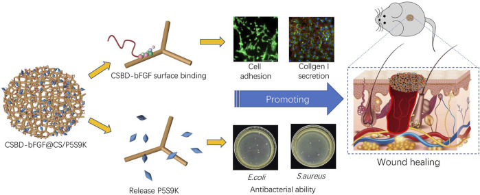
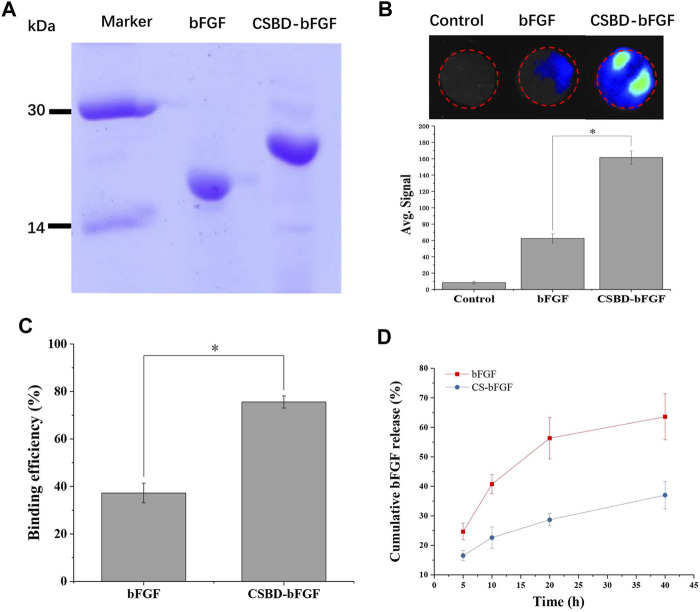
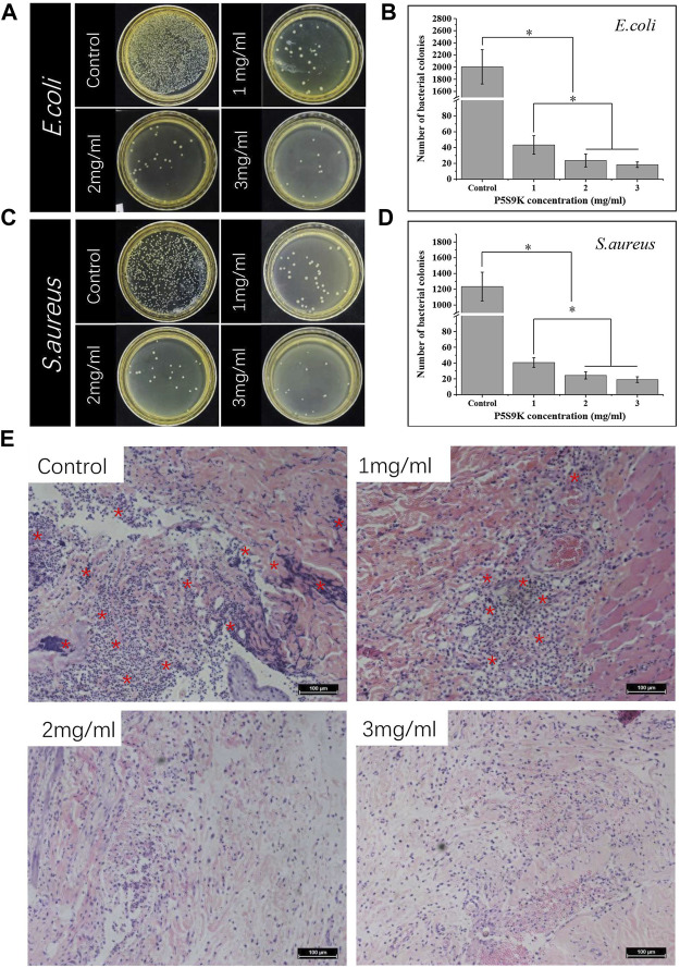
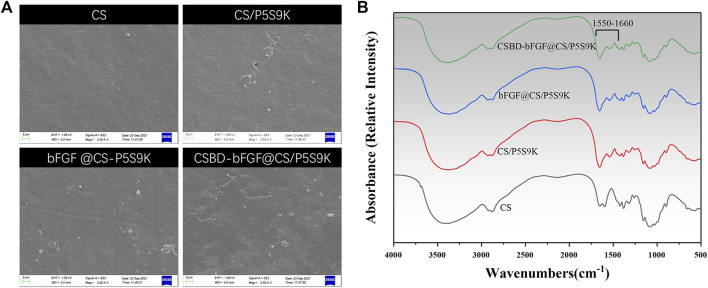
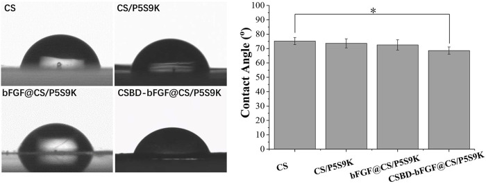
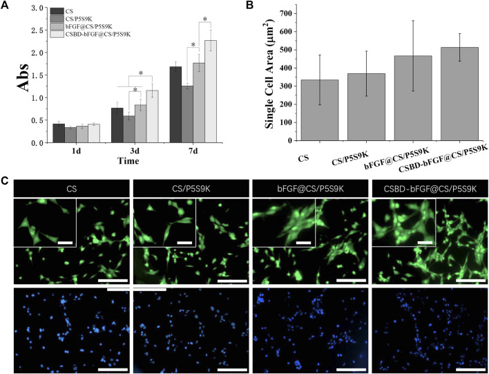
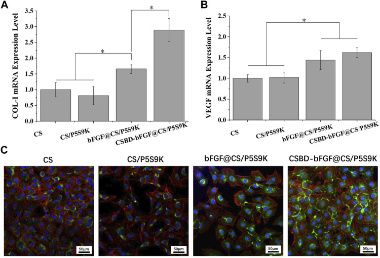
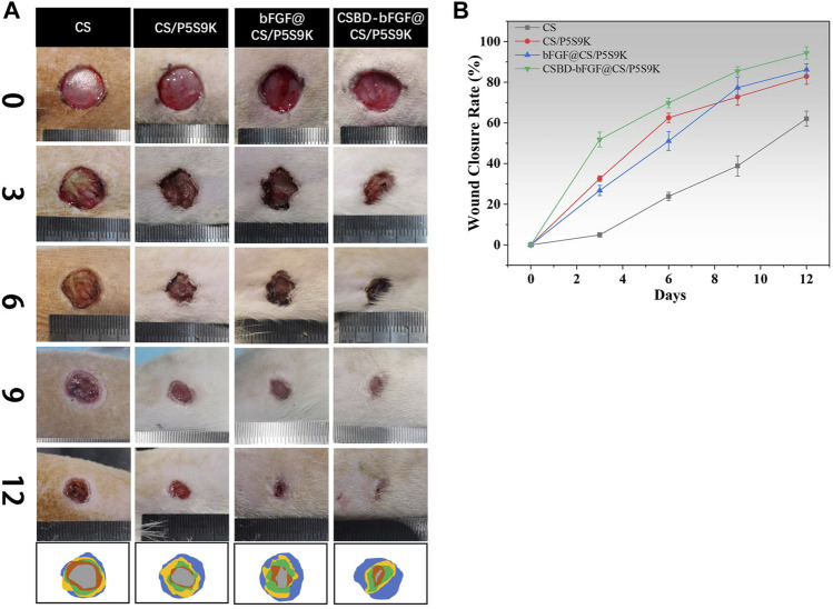
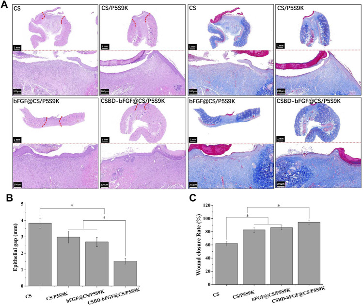
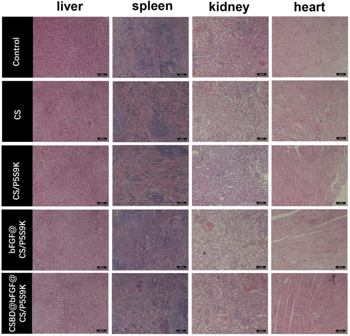
Skin wound healing is an important clinical challenge, and the main treatment points are accelerating epidermal regeneration and preventing infection. Therefore, it is necessary to develop a wound dressing that can simultaneously cure bacterial infections and accelerate wound healing. Here, we report a multifunctional composite wound dressing loaded with chitosan (CS)-binding bFGF (CSBD-bFGF) and antimicrobial peptides (P5S9K). First, CS was used as the dressing matrix material, and P5S9K was encapsulated in CS. Then, CSBD-bFGF was designed by combining recombinant DNA technology and tyrosinase treatment and modified on the dressing material surface. The results show that the binding ability of CSBD-bFGF and CS was significantly improved compared with that of commercial bFGF, and CSBD-bFGF could be controllably released from the CS dressing. More importantly, the prepared dressing material showed excellent antibacterial activity in vivo and in vitro and could effectively inhibit the growth of E. coli and S. aureus. Using NIH3T3 cells as cellular models, the results showed that the CSBD-bFGF@CS/P5S9K composite dressing was a friendly material for cell growth. After cells were seeded on the composite dressing surface, collagen-1 (COL-1) and vascular endothelial growth factor (VEGF) genes expression in cells were significantly upregulated. Finally, the full-thickness wound of the rat dorsal model was applied to analyse the tissue repair ability of the composite dressing. The results showed that the composite dressing containing CSBD-bFGF and P5S9K had the strongest ability to repair skin wounds. Therefore, the CSBD-bFGF@CS/P5S9K composite dressing has good antibacterial and accelerated wound healing abilities and has good application prospects in the treatment of skin wounds.
Keywords: CSBD-bFGF; P5S9K; antimicrobial peptides; chitosan; skin dressing; skin wound.
Copyright © 2022 Hou, Wang, Wang and Song.
Conflict of interest statement
The authors declare that the research was conducted in the absence of any commercial or financial relationships that could be construed as a potential conflict of interest.
Figures
Similar articles
-
Enhanced wound repair ability of arginine-chitosan nanocomposite membrane through the antimicrobial peptides-loaded polydopamine-modified graphene oxide.J Biol Eng. 2021 May 22;15(1):17. doi: 10.1186/s13036-021-00268-3. J Biol Eng. 2021. PMID: 34022941 Free PMC article.
-
Carboxymethyl chitosan-based hydrogels containing fibroblast growth factors for triggering diabetic wound healing.Carbohydr Polym. 2022 Jul 1;287:119336. doi: 10.1016/j.carbpol.2022.119336. Epub 2022 Mar 11. Carbohydr Polym. 2022. PMID: 35422300
-
Chitosan-based carbon nitride-polydopamine‑silver composite dressing with antibacterial properties for wound healing.Carbohydr Polym. 2023 Mar 1;303:120436. doi: 10.1016/j.carbpol.2022.120436. Epub 2022 Dec 9. Carbohydr Polym. 2023. PMID: 36657833
-
Chitosan-Based Dressing Materials for Problematic Wound Management.Adv Exp Med Biol. 2018;1077:527-537. doi: 10.1007/978-981-13-0947-2_28. Adv Exp Med Biol. 2018. PMID: 30357707 Review.
-
A cooling wound dressing for accelerating healing under sunlight.Innovation (Camb). 2024 Jul 2;5(5):100670. doi: 10.1016/j.xinn.2024.100670. eCollection 2024 Sep 9. Innovation (Camb). 2024. PMID: 39108618 Free PMC article. Review. No abstract available.
Cited by
-
Progress in Wound-Healing Products Based on Natural Compounds, Stem Cells, and MicroRNA-Based Biopolymers in the European, USA, and Asian Markets: Opportunities, Barriers, and Regulatory Issues.Polymers (Basel). 2024 May 3;16(9):1280. doi: 10.3390/polym16091280. Polymers (Basel). 2024. PMID: 38732749 Free PMC article. Review.
-
Bioactive peptides and proteins for tissue repair: microenvironment modulation, rational delivery, and clinical potential.Mil Med Res. 2024 Dec 5;11(1):75. doi: 10.1186/s40779-024-00576-x. Mil Med Res. 2024. PMID: 39639374 Free PMC article. Review.
-
Nanoscale borosilicate bioactive glass for regenerative therapy of full-thickness skin defects in rabbit animal model.Front Bioeng Biotechnol. 2023 May 18;11:1036125. doi: 10.3389/fbioe.2023.1036125. eCollection 2023. Front Bioeng Biotechnol. 2023. PMID: 37274157 Free PMC article.
-
Polydopamine-coated polycaprolactone/carbon nanotube fibrous scaffolds loaded with basic fibroblast growth factor for wound healing.Mater Today Bio. 2024 Aug 6;28:101190. doi: 10.1016/j.mtbio.2024.101190. eCollection 2024 Oct. Mater Today Bio. 2024. PMID: 39221197 Free PMC article.
-
Fabrication of bFGF/polydopamine-loaded PEEK implants for improving soft tissue integration by upregulating Wnt/β-catenin signaling.Heliyon. 2023 Mar 21;9(4):e14800. doi: 10.1016/j.heliyon.2023.e14800. eCollection 2023 Apr. Heliyon. 2023. PMID: 37012909 Free PMC article.
References
-
- Akasaka Y., Fujisawa C., Nakamichi M., Okaneya T. (2020). bFGF Induced Angiogenesis in Wounds. Wound Repair Regen. 28 (5), A3. 10.1111/wrr.12843 - DOI
-
- Khan Z. A., Jamil S., Akhtar A., Mustehsan Bashir M., Yar M. (2020). Chitosan Based Hybrid Materials Used for Wound Healing Applications- A Short Review. Int. J. Polymeric Mater. Polymeric Biomater. 69 (7), 419–436. 10.1080/00914037.2019.1575828 - DOI
-
- Bakhsheshi-Rad H. R., Ismail A. F., Aziz M., Akbari M., Hadisi Z., Omidi M., et al. (2020). Development of the PVA/CS Nanofibers Containing Silk Protein Sericin as a Wound Dressing: In Vitro and In Vivo Assessment. Int. J. Biol. Macromolecules 149, 513–521. 10.1016/j.ijbiomac.2020.01.139 - DOI - PubMed
LinkOut - more resources
Full Text Sources