Review
. 2022 Apr 25:13:781063.
doi: 10.3389/fneur.2022.781063. eCollection 2022.

Monocarboxylate Transporter 1 May Benefit Cerebral Ischemia via Facilitating Lactate Transport From Glial Cells to Neurons

Affiliations
Review

Monocarboxylate Transporter 1 May Benefit Cerebral Ischemia via Facilitating Lactate Transport From Glial Cells to Neurons

Mao Zhang et al. Front Neurol. .

Abstract

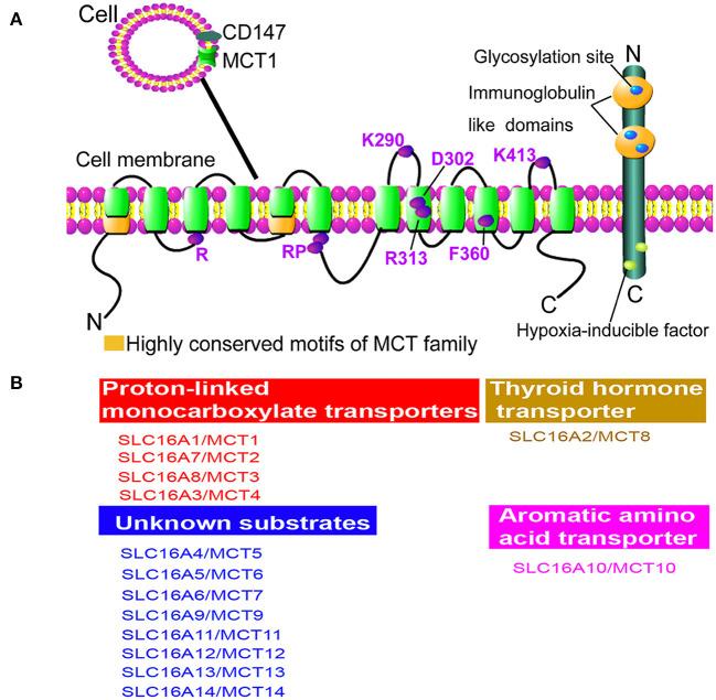
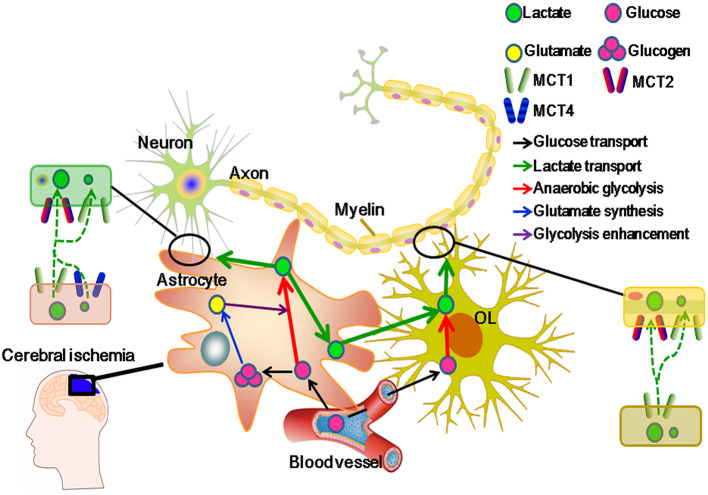
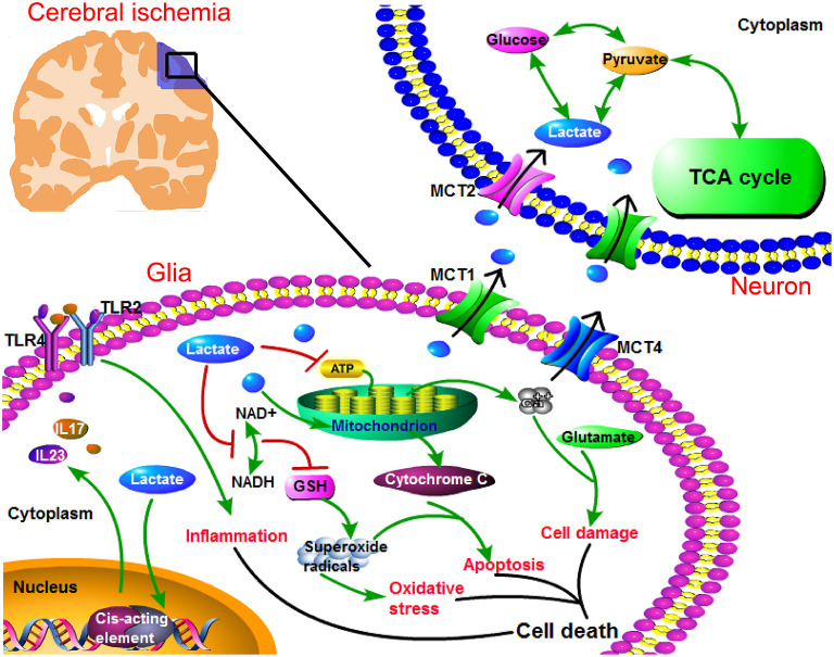
Monocarboxylate transporter 1 (MCT1) is expressed in glial cells and some populations of neurons. MCT1 facilitates astrocytes or oligodendrocytes (OLs) in the energy supplement of neurons, which is crucial for maintaining the neuronal activity and axonal function. It is suggested that MCT1 upregulation in cerebral ischemia is protective to ischemia/reperfusion (I/R) injury. Otherwise, its underlying mechanism has not been clearly discussed. In this review, it provides a novel insight that MCT1 may protect brain from I/R injury via facilitating lactate transport from glial cells (such as, astrocytes and OLs) to neurons. It extensively discusses (1) the structure and localization of MCT1; (2) the regulation of MCT1 in lactate transport among astrocytes, OLs, and neurons; and (3) the regulation of MCT1 in the cellular response of lactate accumulation under ischemic attack. At last, this review concludes that MCT1, in cerebral ischemia, may improve lactate transport from glial cells to neurons, which subsequently alleviates cellular damage induced by lactate accumulation (mostly in glial cells), and meets the energy metabolism of neurons.

Keywords: astrocytes; cerebral ischemia; lactate transport; monocarboxylate transporter 1 (MCT1); oligodendrocytes (OLs).


Conflict of interest statement

The authors declare that the research was conducted in the absence of any commercial or financial relationships that could be construed as a potential conflict of interest.

Figures

Figure 1
Figure 1
Structure and function of monocarboxylate transporter (MCT) family. (A) The proposed topology of MCT family. There are 10–12 α-helical transmembrane domains (TMDs) for MCT family membranes in the presence of N- and C-termini located within cytoplasm. Besides, it shows the greatest variation at N- and C-termini and the large loop between TMDs 6 and 7 among family members, while the TMDs themselves are highly conserved. Importantly, two highly conserved motifs can be identified as the characteristic of the MCT family (green). Cluster of differentiation 147 (CD147) is an ancillary protein of MCT1 and MCT4, which is a cell surface glycoprotein containing a signal transmembrane span, two immunoglobulin-like domains in the extracellular region, and a short C-terminal cytoplasmic tail. The marked amino acids are significant for the structure conservation of MCT family. Specifically, R represents Arg; RP represents Arg and Pro; K290 represents Lys290; D302 represents Asp302; R313 represents Arg313; F360 represents Phe360; K413 represents Lys413; E218 represents Glu218. (B) Functions of MCT family members. MCT1, MCT2, MCT3, and MCT4 are proton-dependent transporters, which transport lactate, pyruvate, D-β-hydroxybutyrate, and acetoacetate (1). MCT8 can transport thyroid hormone and MCT10 is a transporter of aromatic amino acids, while the substrates of other MCT members are still unknown.
Figure 2
Figure 2
Monocarboxylate transporter 1 (MCT1) regulatively facilitates lactate transport from glial cells to neurons in cerebral ischemia. MCT1 is expressed in glial cells, such as astrocytes and oligodendrocytes (OLs), and some populations of neurons in the areas of hippocampus, brainstem, trigeminal ganglion, and cerebellar Purkinje. Meanwhile, another three lactate transporters (MCT2, MCT3, and MCT4) are restricted to cell types: MCT2 in neurons, MCT3 in epithelial cells, and MCT4 in astrocytes. Moreover, MCT1 can regulate lactate influx and efflux among neural cells. MCT2 can merely regulate lactate influx and MCT4 mainly functions in lactate efflux from neural cells. In cerebral ischemia, MCT1 upregulation protects neurons from I/R injury through regulating lactate transport (2). Generally, glucose is transported from capillaries to glial cells, and then to be polymerized into glucogen (stored in astrocytes) or aerobically metabolized. Otherwise, increased glutamate is released from neurons and activates glial glycolysis in cerebral ischemia. As a result, glucose is metabolized into lactate in glial cells. At this moment, lactate becomes the main energetic substrate of neurons. To adapt the changed microenvironment, e.g., increased acidosis, MCT1 activity is strengthened, which not only facilitates the accumulated lactate efflux from glial cells, but also improves the lactate influx to neurons (11).
Figure 3
Figure 3
Regulation of MCT1 in lactate transport promotes neuronal energy supplement, and suppresses cellular damage under ischemic attack. In ischemic brain, anaerobic glycolysis is largely enhanced and leads to lactate overproduction. Lactate becomes the main energy resource for neurons, while its intracellular accumulation (in glial cells) leads to toxic response. Specifically, lactate accumulation associates with cellular responses, such as inflammation, oxidative stress, apoptosis, Ca2+ overload, and glutamate-induced excitotoxicity. Especially, lactate can promote the transcription of proinflammatory factors (interleukin 23/interleukin 17 (IL23/IL17)), which triggers inflammation via toll-like receptor 2/toll-like receptor 4 (TLR2/TLR4). In addition, lactate can suppress the production of glutathione (GSH) which is an indispensable anti-oxidative enzyme, and GSH deficit leads to the increased superoxide radicals. Then, superoxide radicals can further activate oxidative stress. Again, superoxide radicals contribute to apoptosis in the presence of mitochondrial cytochrome C. Furthermore, lactate decreases adenosine triphosphate (ATP) level, resulting in Ca2+ overload. Ca2+ overload coexists with glutamate-induced excitotoxicity which leads to cellular damage. Collectively, intracellular lactate accumulation contributes to these cellular responses, leading to cell death. Therefore, a timely and rightly utilization of lactate protects brain from I/R injury. MCT1 can facilitate lactate efflux from glial cells, and assist lactate influx into neurons. Besides, neuronal MCT2 assists lactate entries into neurons, and then lactate is efficiently utilized by neurons.

References

    1. Zhao Q, Wang S, Li Y, Wang P, Li S, Guo Y, et al. The role of the mitochondrial calcium uniporter in cerebral ischemia/reperfusion injury in rats involves regulation of mitochondrial energy metabolism. Mol Med Rep. (2013) 7:1073–80. 10.3892/mmr.2013.1321 - DOI - PubMed
    1. Deng HJ, Zhang S, Ge HF, Liu L, Liu LT, Feng H, et al. The effect of cyclosporin a on ischemia-reperfusion damage in a mouse model of ischemic stroke. Neurol Res. (2020) 42:721–9. 10.1080/01616412.2020.1762353 - DOI - PubMed
    1. Castillo X, Rosafio K, Wyss MT, Drandarov K, Buck A, Pellerin L, et al. A probable dual mode of action for both L- and D-lactate neuroprotection in cerebral ischemia. J Cereb Blood Flow Metab. (2015) 35:1561–9. 10.1038/jcbfm.2015.115 - DOI - PMC - PubMed
    1. Tassinari ID, Andrade MKG, Rosa LAD, Hoff MLM, Nunes RR, Vogt EL, et al. Lactate administration reduces brain injury and ameliorates behavioral outcomes following neonatal hypoxia-ischemia. Neuroscience. (2020) 448:191–205. 10.1016/j.neuroscience.2020.09.006 - DOI - PubMed
    1. Roumes H, Dumont U, Sanchez S, Mazuel L, Blanc J, Raffard G, et al. Neuroprotective role of lactate in rat neonatal hypoxia-ischemia. J Cereb Blood Flow Metab. (2021) 41:342–58. 10.1177/0271678X20908355 - DOI - PMC - PubMed

LinkOut - more resources