. 2022 Apr 25:9:867136.
doi: 10.3389/fmolb.2022.867136. eCollection 2022.

Small Things Matter: The 11.6-kDa TraB Protein is Crucial for Antibiotic Resistance Transfer Among Enterococci

Affiliations

Small Things Matter: The 11.6-kDa TraB Protein is Crucial for Antibiotic Resistance Transfer Among Enterococci

Tamara M I Berger et al. Front Mol Biosci. .

Abstract

Conjugative transfer is the most important means for spreading antibiotic resistance genes. It is used by Gram-positive and Gram-negative bacteria, and archaea as well. Conjugative transfer is mediated by molecular membrane-spanning nanomachines, so called Type 4 Secretion Systems (T4SS). The T4SS of the broad-host-range inc18-plasmid pIP501 is organized in a single operon encoding 15 putative transfer proteins. pIP501 was originally isolated from a clinical Streptococcus agalactiae strain but is mainly found in Enterococci. In this study, we demonstrate that the small transmembrane protein TraB is essential for pIP501 transfer. Complementation of a markerless pIP501∆traB knockout by traB lacking its secretion signal sequence did not fully restore conjugative transfer. Pull-downs with Strep-tagged TraB demonstrated interactions of TraB with the putative mating pair formation proteins, TraF, TraH, TraK, TraM, and with the lytic transglycosylase TraG. As TraB is the only putative mating pair formation complex protein containing a secretion signal sequence, we speculate on its role as T4SS recruitment factor. Moreover, structural features of TraB and TraB orthologs are presented, making an essential role of TraB-like proteins in antibiotic resistance transfer among Firmicutes likely.

Keywords: Enterococcus; antibiotic resistance; conjugative transfer; plasmid; secretion complex; type IV secretion system.


Conflict of interest statement

The authors declare that the research was conducted in the absence of any commercial or financial relationships that could be construed as a potential conflict of interest.

Figures

FIGURE 1
FIGURE 1
Results of biparental mating assays of pIP501 and derivatives. E. faecalis JH2-2 isogenic wild type (pIP501), mutant strain E. faecalis JH2-2 (pIP501∆traB) and complementation strain where traB, traB with a C-terminal Strep-tag or traB 31-110 are supplied in trans (E. faecalis JH2-2 (pIP501∆traB; pEU327-RBS-traB), E. faecalis JH2-2 (pIP501∆traB; pEU327-RBS-traB-Strep) or E. faecalis JH2-2 (pIP501∆traB; pEU327-RBS-traB 31-110) were applied as donors and E. faecalis OG1X as recipient. Transfer frequencies are presented as the number of transconjugants per recipient cell. n = 4. Mean values are depicted with the standard deviation. ***p < 0.0003, **p < 0.0038 as determined by Welch’s t test. The dashed line depicts the detection limit of the assay (4.4 × 10⁻⁸ transconjugants per recipient).
FIGURE 2
FIGURE 2
(A) Domain composition of TraB. The first predicted helix is part of the secretion signal sequence (predicted with SignalP5.0) including a cleavage site between alanine 30 and alanine 31. Secondary structure elements were calculated with PSIPRED. After a short coil structure, the first transmembrane helix (TMH1) spans from proline 34 to proline 70 followed by a short loop region leading to the second transmembrane helix (TMH2) starting at proline 75 and ending at leucine 109. MEMPACK and MEMSAT-SVM were used to determine the orientation of TraB in the membrane as represented by the color code above the TMHs. Green parts are predicted to be extracellular, golden parts within the membrane and the lavender part intracellular. (B) Multiple sequence alignments using PSI/TM-Coffee for membrane proteins from the T-coffee server of TraB and its orthologs from other putative conjugative systems. Orthologs were identified by an iterative PSI-BLAST using the NCBI databank. Only proteins originating from a conjugative context were included in the multiple sequence alignments. The color scheme shows the orientation of the proteins mapped on to their sequence. The panel to the right shows the evaluation scores from the TCS evaluation. Asterisks “*” show conserved amino acids in all sequences. Colons “:” indicate amino acids of high similarities. Dots “.” indicate partial similarities. In addition, identical and highly similar amino acids are highlighted with grey shading of the respective alignment boxes. The numbers above the aligned sequences are referring to the amino acid position of TraBpIP501 (C) Western Blot of the cell fractionation of E. faecalis JH2-2 harboring different plasmids using a specific antibody against TraBpIP501. The plasmids are given below the membrane. The fractions are labeled as: CW: cell wall; CM: cell membrane; CP: cytoplasm. TraB (white arrow heads) shows an unusual migration behavior with an apparent MW of 20 kDa on the SDS-PAGE, which is commonly observed for membrane proteins and may be explained by the atypical SDS binding (Rath et al., 2009).
FIGURE 3
FIGURE 3
(A) Alignment of TraBpIP501 and its orthologs using the theoretical structures from Robetta. TrsBpAMβ1 and TrsBpRE25 were not included, because both are 100% identical to TraBpIP501. TraBpIP501 is shown in orange; the mating channel formation protein from pUCB11B is shown in purple; the CagC family protein from Staphylococcus aureus AF2236 is shown in rose; the CagC family protein from Clostridium algoriphilum DSM 16153 is depicted in green; TraB from Staphylococcus aureus 3688STDY6124879 is shown in slate; TrsBpV030-8 is depicted in green-blue. (B) Shows TraBpIP501 colored according to its orientation in the cell membrane. The grey part represents the secretion signal sequence; slate parts the extracellular part; the salmon parts are predicted to be within the membrane; the yellow parts are cytosolic. Amino acids, which are conserved in the multiple sequence alignments are shown in stick presentation. (C) and (D) Zoom into the areas of TraBpIP501 showing the highest conservation. The conserved amino acids are shown as sticks and labeled. Same color scheme as in panel (B,C) Showing the details of the interface area between the cell membrane and the peptidoglycan layer. (D) Zoom into the cytosolic loop of TraBpIP501, bottom view.
FIGURE 4
FIGURE 4
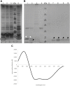
(A) Silver-stained 12% SDS polyacrylamide gel of the elution fractions from IMAC of the TraB31-110 test purification with two different E. coli expression strains. For lane 1-3 BL21 Codon Plus RIL cells were used, for lane 5 BL21 star cells. Lane 4 shows the MW standard (Thermo Fisher Scientific). Circled bands were cut out and applied to MS analysis (B) Coomassie-stained 12% SDS polyacrylamide gel showing different purification steps of TraB31-110. Lane 1: pooled elution fraction of the IMAC; lane 2: same fractions after TEV cleavage; lane 3 and 4: elution fractions of the reverse IMAC; lane 5: MW standard (Thermo Fisher Scientific); lane 6–10: elution fractions of the SEC using a Superdex 200 increase column. White arrow heads point at uncleaved TraB31-110. Black arrow heads point at TEV-cleaved TraB31-110. (C) CD spectrum of TraB31-110 showing two minima at 209 and 222 nm implying a mainly α-helical conformation of TraB31-110.
FIGURE 5
FIGURE 5
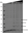
12% SDS polyacrylamide gel of in vitro cross-linking of TraB31-110 using increasing glutaraldehyde concentrations (0/0.001/0.01/0.05/0.1%) from left to right. Lane 1 shows the MW standard (Thermo Fisher Scientific).
FIGURE 6
FIGURE 6
Western Blot of the elution fractions of the pull-down with TraB-Strep. The used anti-Tra antibody and the MW of the respective TrapIP501 protein is given above the blot. The TraG protein runs significantly faster than its calculated MW. One possible explanation would be that only the CHAP domain of TraG is part of the eluted protein complex. The two TraK bands correspond to its two start codons resulting in proteins of sizes of 36.4 and 32.3 kDa. The Western Blot with anti-TraO antibody was performed as negative control. TraO is present in the lysate (lane 1) and in the flow through of the column (lane 2), but not in the elution fraction (lane 3). The two bands are showing TraO with and without its secretion sequence (the difference in MW = 2.5 kDa). The slower migration of the TraO bands may be explained by the high acidity of the proteins (pI 3.8 and 3.7) (Matagne et al., 1991).

References

    1. Abajy M. Y., Kopeć J., Schiwon K., Burzynski M., Döring M., Bohn C., et al. (2007). A Type IV-secretion-like System Is Required for Conjugative DNA Transport of Broad-Host-Range Plasmid pIP501 in Gram-Positive Bacteria. J. Bacteriol. 189, 2487–2496. 10.1128/JB.01491-06 - DOI - PMC - PubMed
    1. Almagro Armenteros J. J., Tsirigos K. D., Sønderby C. K., Petersen T. N., Winther O., Brunak S., et al. (2019). Signalp 5.0 Improves Signal Peptide Predictions Using Deep Neural Networks. Nat. Biotechnol. 37, 420–423. 10.1038/s41587-019-0036-z - DOI - PubMed
    1. Alvarez-Martinez C. E., Christie P. J. (2009). Biological Diversity of Prokaryotic Type IV Secretion Systems. Microbiol. Mol. Biol. Rev. 73, 775–808. 10.1128/MMBR.00023-09 - DOI - PMC - PubMed
    1. Arends K., Celik E.-K., Probst I., Goessweiner-Mohr N., Fercher C., Grumet L., et al. (2013). TraG Encoded by the pIP501 Type IV Secretion System Is a Two-Domain Peptidoglycan-Degrading Enzyme Essential for Conjugative Transfer. J. Bacteriol. 195, 4436–4444. 10.1128/JB.02263-12 - DOI - PMC - PubMed
    1. Baek M., DiMaio F., Anishchenko I., Dauparas J., Ovchinnikov S., Lee G. R., et al. (2021). Accurate Prediction of Protein Structures and Interactions Using a Three-Track Neural Network. Science 373, 871–876. 10.1126/SCIENCE.ABJ8754 - DOI - PMC - PubMed

LinkOut - more resources