

Development of a visible light, cross-linked GelMA hydrogel containing decellularized human amniotic particles as a soft tissue replacement for oral mucosa repair
- PMID: 35547651
- PMCID: PMC9087906
- DOI: 10.1039/c9ra03009c
Development of a visible light, cross-linked GelMA hydrogel containing decellularized human amniotic particles as a soft tissue replacement for oral mucosa repair
Abstract
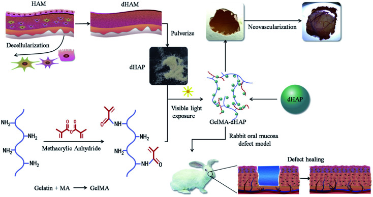
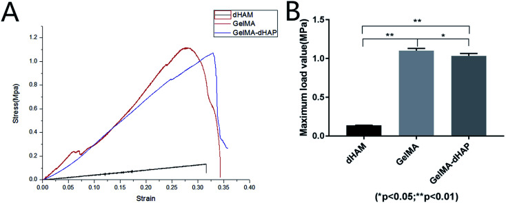
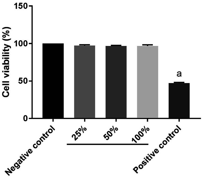
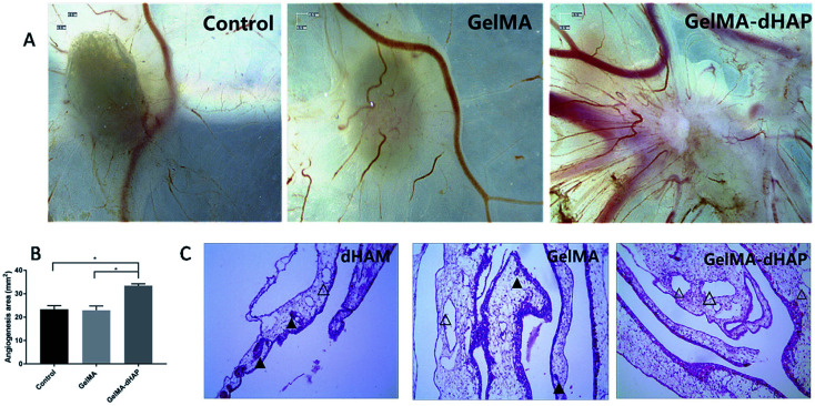
Early effective treatment of oral mucosal defects is the key to ensuring defect healing and functional recovery. The application of human amniotic membrane (HAM) in promoting wound healing has been shown to be safe and effective. However, amniotic membrane is thin, easy to tear and difficult to handle. Combined with the natural forces at play in the oral cavity, this has restricted the clinical applications of HAM for healing of mucosal defects. Methacrylated gelatin (GelMA) has good mechanical strength and adhesion, and can be used as a bionic repair film to attach to the damaged surface of oral mucosa, but GelMA lacks bioactive substances and cannot promote the rapid repair of oral mucosal defects. The aim of this study was to design a type of composite GelMA hydrogel mixed with decellularized human amniotic particles (dHAP) as an oral mucosa substitute, to promote regeneration of defective mucosa by stimulating rapid angiogenesis. The composite substitute GelMA-dHAP was easy to synthesize and store, and easy to operate for repair of oral mucosal defects. We show the angiogenic potential of GelMA-dHAP on chick chorioallontoic membrane and the curative effect of GelMA-dHAP as a treatment in the rabbit oral mucosa defect model. In conclusion, this study confirms the effectiveness of GelMA-dHAP as an ideal soft tissue substitute for the repair of oral mucosal defects, overcoming the shortcomings of using HAM or GelMA alone.
This journal is © The Royal Society of Chemistry.
Conflict of interest statement
The authors declared that there was no conflict of interest in the financial and publication of the work. All the authors gave their seal of approval to the manuscript.
Figures

Similar articles
-
Photo-crosslinkable amniotic membrane hydrogel for skin defect healing.Acta Biomater. 2021 Apr 15;125:197-207. doi: 10.1016/j.actbio.2021.02.043. Epub 2021 Mar 4. Acta Biomater. 2021. PMID: 33676048
-
Design and fabrication of gelatin-based hydrogel loaded with modified amniotic extracellular matrix for enhanced wound healing.Heliyon. 2023 Sep 28;9(10):e20521. doi: 10.1016/j.heliyon.2023.e20521. eCollection 2023 Oct. Heliyon. 2023. PMID: 37790967 Free PMC article.
-
Biohybrid methacrylated gelatin/polyacrylamide hydrogels for cartilage repair.J Mater Chem B. 2017 Jan 28;5(4):731-741. doi: 10.1039/c6tb02348g. Epub 2017 Jan 3. J Mater Chem B. 2017. PMID: 32263841
-
Recent Advances on Bioprinted Gelatin Methacrylate-Based Hydrogels for Tissue Repair.Tissue Eng Part A. 2021 Jun;27(11-12):679-702. doi: 10.1089/ten.TEA.2020.0350. Epub 2021 Mar 9. Tissue Eng Part A. 2021. PMID: 33499750 Review.
-
Introduction to Amniotic Membranes in Maxillofacial Surgery-A Scoping Review.Medicina (Kaunas). 2024 Apr 19;60(4):663. doi: 10.3390/medicina60040663. Medicina (Kaunas). 2024. PMID: 38674309 Free PMC article.
Cited by
-
Applications of Gelatin Methacryloyl (GelMA) Hydrogels in Microfluidic Technique-Assisted Tissue Engineering.Molecules. 2020 Nov 13;25(22):5305. doi: 10.3390/molecules25225305. Molecules. 2020. PMID: 33202954 Free PMC article. Review.
-
A comparative study of human amniotic membrane, tilapia skin collagen, and Centella asiatica derived gel to treat burn wound in rat model.Cell Tissue Bank. 2025 Jan 16;26(1):8. doi: 10.1007/s10561-025-10157-4. Cell Tissue Bank. 2025. PMID: 39821851
-
Applications of the amniotic membrane in tissue engineering and regeneration: the hundred-year challenge.Stem Cell Res Ther. 2022 Jan 10;13(1):8. doi: 10.1186/s13287-021-02684-0. Stem Cell Res Ther. 2022. PMID: 35012669 Free PMC article. Review.
-
Dehydrated Human Amnion-Chorion Membrane as a Bioactive Scaffold for Dental Pulp Tissue Regeneration.Biomimetics (Basel). 2024 Dec 18;9(12):771. doi: 10.3390/biomimetics9120771. Biomimetics (Basel). 2024. PMID: 39727775 Free PMC article. Review.
-
Snake extract-laden hemostatic bioadhesive gel cross-linked by visible light.Sci Adv. 2021 Jul 14;7(29):eabf9635. doi: 10.1126/sciadv.abf9635. Print 2021 Jul. Sci Adv. 2021. PMID: 34261653 Free PMC article.
References
-
- Piraino F. Selimovic S. Mol. Cell. Ther. 2015;2015:1–10.
-
- Rigal-Satourne J. C. Legeais J. M. Texier J. M. Savoldelli M. Renard J. P. Maurin J. F. Renard G. Invest. Ophthalmol. Visual Sci. 2000;4:S455.
LinkOut - more resources
Full Text Sources
Other Literature Sources