. 2018 Sep 21;8(57):32621-32636.
doi: 10.1039/c8ra06362a. eCollection 2018 Sep 18.

Formulation, characterization and evaluation of morusin loaded niosomes for potentiation of anticancer therapy

Affiliations

Formulation, characterization and evaluation of morusin loaded niosomes for potentiation of anticancer therapy

Srishti Agarwal et al. RSC Adv. .

Abstract

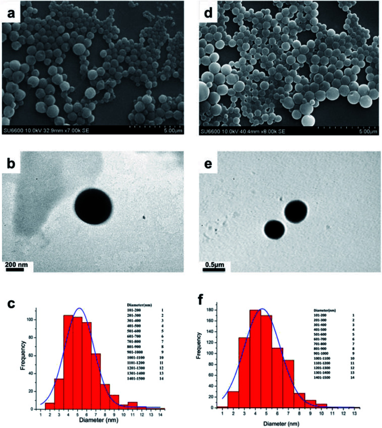
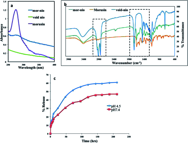
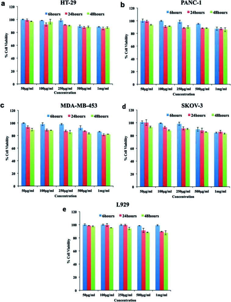
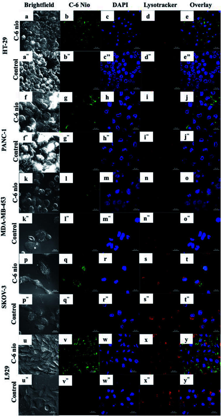
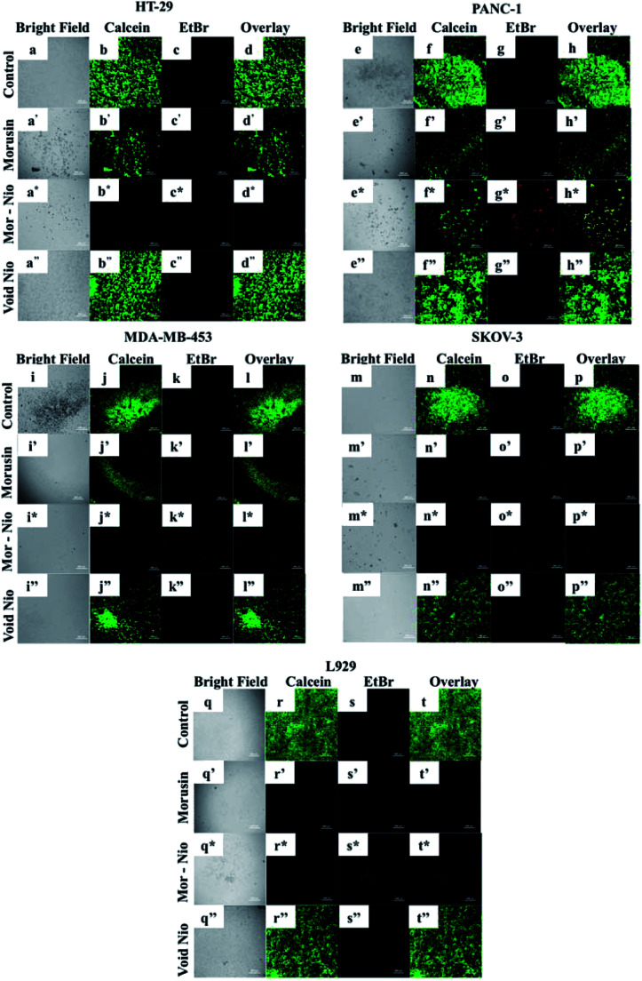
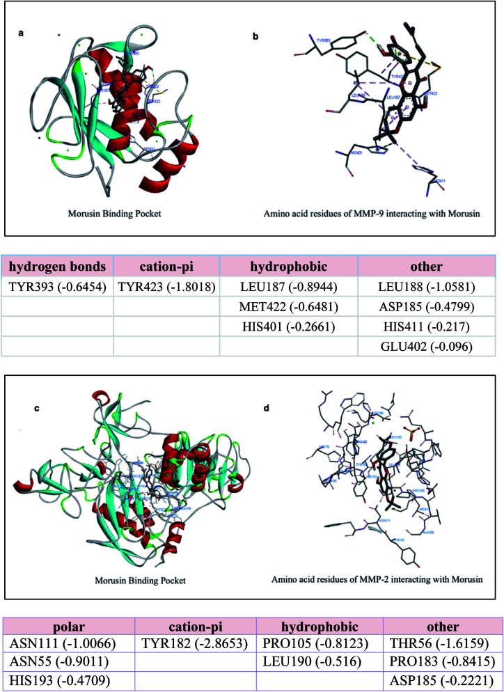
Morusin, a water-insoluble prenylated flavonoid is known for its numerous medicinal properties. It manifests its anticancer potential by suppression of genes involved in tumor progression. However, poor solubility of the drug results in low bioavailability and rapid degradation thus hindering its clinical utilization. In order to overcome this, we have synthesized a niosome system composed of non-ionic surfactant span 60 and cholesterol using a thin-layer evaporation technique to improve the aqueous-phase solubility of the drug. Highly cytocompatible niosomes of 479 nm average size with smooth and uniform spherical morphology were synthesized in a facile manner. Unlike free morusin, nanomorusin was found to be freely dispersible in aqueous media. Having an extremely high drug entrapment efficiency (97%), controlled and sustained release of morusin resulting in enhanced therapeutic efficacy was observed in cancer cell lines of 4 different lineages. The results demonstrate that the morusin-niosome system is a promising strategy for enhanced anti-cancer activity against multiple cancer types and could be an indispensable tool for future targeted chemotherapeutic strategies.


Conflict of interest statement

There are no conflicts to declare.

Figures

Fig. 1
Fig. 1. (a) SEM micrograph of void nio (d) SEM micrograph of mor-nio. (b) TEM image of void nio (e) TEM image of mor-nio (c and f) Gaussian size distribution for void and mor-nio.
Fig. 2
Fig. 2. (a) UV-absorption overlay spectra for free morusin, mor-nio and void nio. The characteristic peak for morusin is around 269 nm, which could be also observed in mor-nio. The void nio provided a flat profile (b) FT-IR spectra of mor-nio in comparison to native morusin and void nio as a control. (c) Sustained drug release profile for morusin nano formulation. An enhanced release was observed in acidic pH establishing mor-nio as an effective drug delivery system for pH dependent dug release in cancer cells.
Fig. 3
Fig. 3. Cytocompatibility of void niosomes tested against cancer (a) HT-29, (b) PANC-1, (c) MDA-MB-453 (d) SKOV-3, cells and normal cell line (e) L929. The particles were highly cytocompatible as depicted by high viability in all cell lines even at highest concentration of particles used.
Fig. 4
Fig. 4. Confocal microscopy images demonstrating cellular internalization of coumarin-6 niosomes by cancer cells. The green fluorescent intensity from cells with C-6 nio was clearly evident, signifying the enhanced uptake of niosomes by cancer cells. Nuclei were stained with NucBlue and lysosomes with lysotracker deep red to show nanoparticles co-localization. Scale bars represent 20 μm.
Fig. 5
Fig. 5. Therapeutic efficacy of morusin-loaded niosomes and free morusin towards (a and b) HT-29 (c and d) PANC-1 (e and f) MDA-MB-453 (g and h) SKOV-3 and (i and j) L929 cell line. Compared to the free drug, more inhibition was observed in the case of morusin-loaded niosomes in all cell lines.
Fig. 6
Fig. 6. Therapeutic efficacy of morusin loaded, free morusin and void niosomes against cancer cell lines using Live/Dead Assay. The control cells and cells treated with void niosomes were live as evident by the green fluorescence. The cells treated with morusin niosomes showed considerable toxicity with minimal number of live cells and increased amount of dead cells (red fluorescence) compared to free morusin where there were both viable and dead cell populations. Scale bars represent 500 μm.
Fig. 7
Fig. 7. Molecular docking studies showing interaction between the metalloproteinases (MMP-9 and MMP-2) and morusin. (a and b) It shows the amino acid residues of MMP-9 interacting with morusin (c and d) Probable binding pocket for morusin on MMP-2 and its interacting residues, which help in attaining stable conformational state via polar and non-polar interactions.

References

    1. Prager G. W. Braga S. Bystricky B. Qvortrup C. Criscitiello C. Esin E. et al. ESMO Open. 2018;3(2):e000285. doi: 10.1136/esmoopen-2017-000285. - DOI - PMC - PubMed
    1. Xie J. Fan Z. Li Y. Zhang Y. Yu F. Su G. Xie L. Hou Z. Int. J. Nanomed. 2018;13:1381–1398. doi: 10.2147/IJN.S152312. - DOI - PMC - PubMed
    1. Davatgaran-Taghipour Y. Masoomzadeh S. Farzaei M. H. Bahramsoltani R. Karimi-Soureh Z. Rahimi R. Abdollahi M. Int. J. Nanomed. 2017;12:2689–2702. doi: 10.2147/IJN.S131973. - DOI - PMC - PubMed
    1. Singh S. K. Lillard Jr. J. W. Singh R. Cancer Lett. 2018;427:49–62. doi: 10.1016/j.canlet.2018.04.017. - DOI - PMC - PubMed
    1. Kamal R. Chadha V. D. Dhawan D. K. Nanomedicine. 2018;14:1059–1071. doi: 10.1016/j.nano.2018.01.008. - DOI - PubMed