. 2022 Apr 25:13:883743.
doi: 10.3389/fimmu.2022.883743. eCollection 2022.

The Hidden Side of Complement Regulator C4BP: Dissection and Evaluation of Its Immunomodulatory Activity

Affiliations

The Hidden Side of Complement Regulator C4BP: Dissection and Evaluation of Its Immunomodulatory Activity

Inmaculada Serrano et al. Front Immunol. .

Abstract

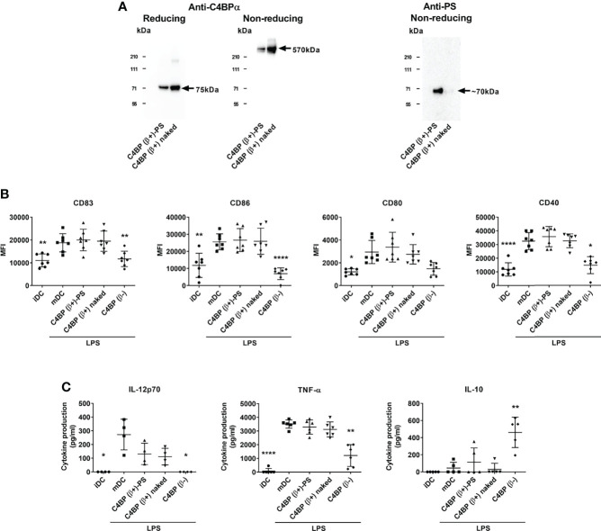
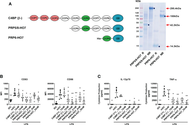
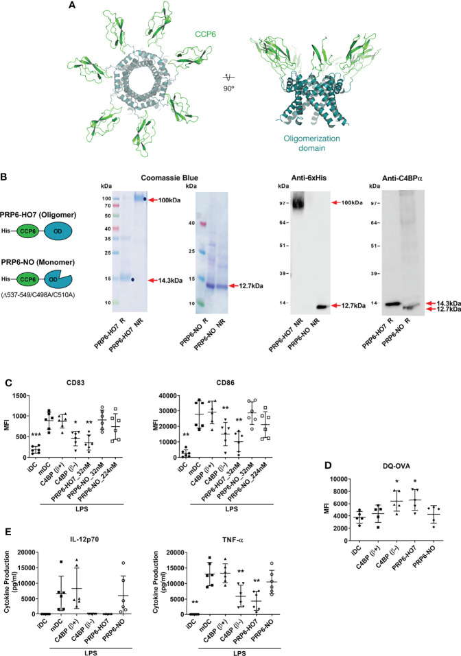
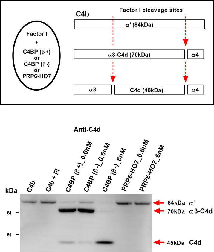
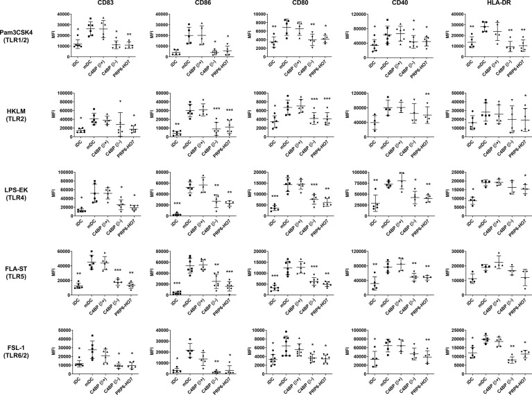
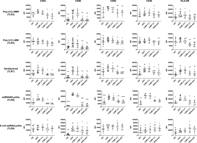
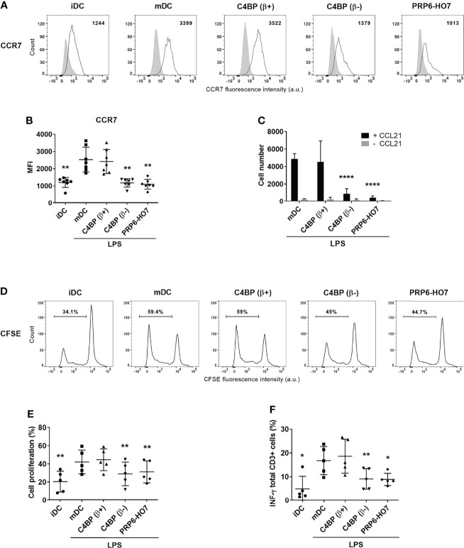
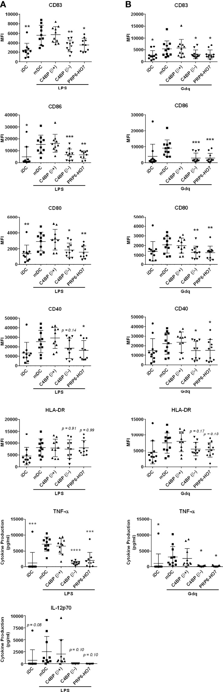
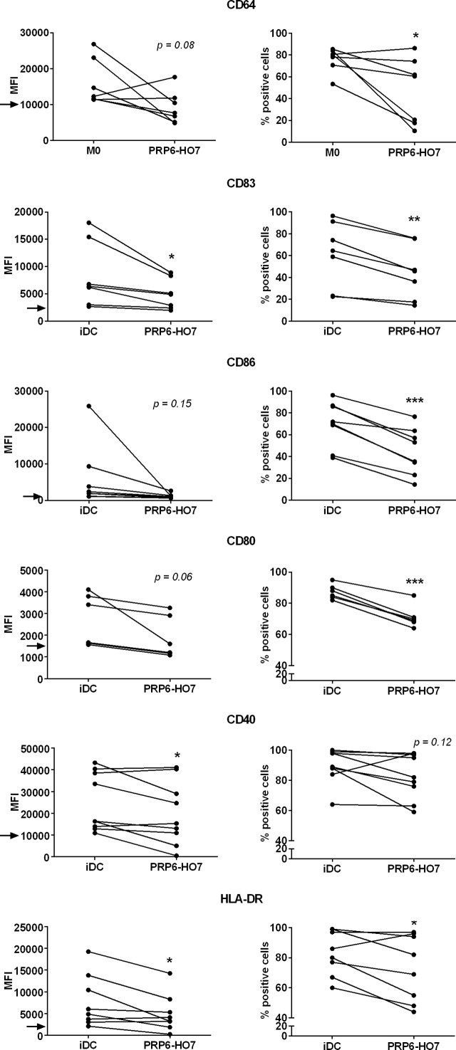
C4b-binding protein (C4BP) is a well-known regulator of the complement system that holds additional and important activities unrelated to complement inhibition. Recently, we have described a novel immunomodulatory activity in the minor C4BP(β-) isoform directly acting over inflammatory phagocytes. Here we show that incorporation of the β-chain to the C4BP α-chain oligomer interferes with this immunomodulatory activity of C4BP. Moreover, an oligomeric form including only the complement control protein 6 (CCP6) domain of the C4BP α-chain (PRP6-HO7) is sufficient to "reprogram" monocyte-derived DCs (Mo-DCs) from a pro-inflammatory and immunogenic phenotype to an anti-inflammatory and tolerogenic state. PRP6-HO7 lacks complement regulatory activity but retains full immunomodulatory activity over inflammatory Mo-DCs induced by TLRs, characterized by downregulation of relevant surface markers such as CD83, HLA-DR, co-stimulatory molecules such as CD86, CD80 and CD40, and pro-inflammatory cytokines such as IL-12 and TNF-α. Furthermore, PRP6-HO7-treated Mo-DCs shows increased endocytosis, significantly reduced CCR7 expression and CCL21-mediated chemotaxis, and prevents T cell alloproliferation. Finally, PRP6-HO7 shows also full immunomodulatory activity over Mo-DCs isolated from lupus nephritis patients with active disease, even without further pro-inflammatory stimulation. Therefore PRP6-HO7, retaining the immunomodulatory activity of C4BP(β-) and lacking its complement regulatory activity, might represent a promising and novel alternative to treat autoimmune diseases.

Keywords: C4BP(β-); PRP6-HO7; dendritic cells; immunomodulation; inflammation; lupus nephritis.


Conflict of interest statement

IS, AL and JA are co-inventors on pending or issued patents involving compounds and methods for immunomodulation. The remaining authors declare that the research was conducted in the absence of any commercial or financial relationships that could be construed as a potential conflict of interest.

Figures

Figure 1
Figure 1
The β-chain of C4BP impairs the immunomodulatory activity of its internal CCP6 α-chain domain. (A) SDS-PAGE and Western analysis of the major physiological C4BP isoform (C4BP(β+)) complexed with PS (C4BP(β+)-PS) through its β-chain, and the same C4BP(β+) isoform devoid of PS (C4BP(β+) naked). Both proteins were resolved under reducing (left) and non-reducing (middle) conditions and probed with PK9008 anti-C4BP α-chain antibody. A further Western blot probed with an anti-PS antibody confirmed the absence of PS in the “C4BP(β+) naked” form (right). (B) Human Mo-DCs were incubated throughout their differentiation process with C4BP(β+)-PS, C4BP(β+) naked and C4BP(β-), all at 12 nM (corresponding to 6.0 μg/ml, 5.3 μg/ml, and 5.0 μg/ml, respectively). DC maturation was achieved by LPS treatment. Cells were then collected, washed, and analyzed by flow cytometry for cell surface expression of the activation marker CD83 and the co-stimulatory molecules CD86, CD80 and CD40. MFI, median fluorescence intensity for the different surface markers. (C) The concentrations of IL-12p70, TNF-α and IL-10 were analyzed in the respective cell supernatants by ELISA. iDC, untreated, immature DCs; mDC, untreated, LPS-matured DCs. The results shown are the mean ± SD from 7 independent donors (cell surface markers), or from 4-6 independent donors performed in duplicate (cytokines) (*p < 0.05; **p < 0.01; ****p < 0.0001 compared with mDC). IL-12p70 concentrations induced by C4BP(β+)-PS and C4BP(β+) naked appeared reduced but were not statistically significant compared to that induced by mDC (p = 0.082, and p = 0.053, respectively).
Figure 2
Figure 2
Deletion of the complement inhibitory domains of C4BP(β-) does not affect its immunomodulatory activity. (A) Left: Schematic structure of the α-chains from C4BP(β−), and from its variants PRP5/8-HO7 and PRP6-HO7. The CCP1-CCP3 complement inhibitory domains are depicted in red. The CCP6 immunomodulatory domain is depicted in green. The oligomerization domain (OD) is depicted in blue. “His” refers to a poly-histidine tag located at the N-terminus of PRP6-HO7. Right: SDS-PAGE and Coomassie Blue staining of both PRP5/8-HO7 and PRP6-HO7 under reducing (R) and non-reducing (NR) conditions. Red arrows and blue dots indicate the location and size of both reduced and non-reduced protein forms. Left lane, molecular weight marker. (B) Human Mo-DCs were incubated throughout their differentiation process with C4BP(β+), C4BP(β-) (both at 12 nM) and the variants PRP5/8-HO7 and PRP6-HO7 at the indicated concentrations. DC maturation was achieved by LPS treatment. Cells were then collected, washed, and analyzed by flow cytometry for cell surface expression of the activation marker CD83 and the co-stimulatory molecule CD86. MFI, median fluorescence intensity for the different surface markers. (C) The concentrations of IL-12p70 and TNF-α were analyzed in the respective cell supernatants by ELISA. iDC, untreated, immature DCs; mDC, untreated, LPS-matured DCs. The results shown are the mean ± SD from 7 independent donors (cell surface markers), or from 5-6 independent donors performed in duplicate (cytokines) (*p < 0.05; **p < 0.01 compared with mDC).
Figure 3
Figure 3
Oligomerization is necessary to preserve the immunomodulatory activity of the C4BP α-chain CCP6 domain. (A) Molecular modeling of PRP6-HO7. PRP6-HO7 homo-oligomer structure prediction by comparative protein structure modeling with MODELLER. PRP6-HO7 is shown in cartoon representation. The N-terminal CCP6 domain and the C-terminal oligomerization domain are shown in green and blue, respectively. (B) Schematic structure of the monomer chain from the PRP6-HO7 heptamer, and from PRP6-NO, unable to oligomerize because of a mutated/truncated OD. PRP6-HO7 and PRP6-NO were visualized by SDS-PAGE and Coomassie Blue staining, and by Western analysis against anti-His and anti-C4BP α-chain antibodies, under both reducing (R) (PRP6-HO7 monomer, 14.3 kDa; PRP6-NO, 12.7 kDa) and non-reducing (NR) (PRP6-HO7 oligomer, 100 kDa; PRP6-NO, 12.7 kDa) conditions. Red arrows indicate the respective molecular weights. (C) Human Mo-DCs were incubated throughout their differentiation process with C4BP(β+), C4BP(β-) (both at 12 nM) and the variants PRP6-HO7 and PRP6-NO (both at 32 nM, unless otherwise stated). DC maturation was achieved by LPS treatment. Cells were then collected, washed, and analyzed by flow cytometry for cell surface expression of the activation marker CD83 and the co-stimulatory molecule CD86. MFI, median fluorescence intensity for the different surface markers. The results shown are the mean ± SD from 6 independent donors (*p < 0.05; **p < 0.01; ***p < 0.001 compared with mDC). (D) Comparative endocytic activity of Mo-DCs was also assessed by flow cytometry, measuring fluorescent DQ-OVA internalization (receptor-mediated endocytosis) at the differentiation stage, after treatment with C4BP(β+), C4BP(β-), PRP6-HO7, and PRP6-NO. Data shown are the mean MFI ± SD from 5 independent experiments (*p < 0.05 compared with iDC). (E) The concentrations of IL-12p70 and TNF-α were analyzed in the cell supernatants from (C), except the PRP6-NO_224 nM sample, by ELISA. iDC, untreated, immature DCs; mDC, untreated, LPS-matured DCs. The results shown are the mean ± SD from 6 independent donors performed in duplicate (**p < 0.01 compared with mDC).
Figure 4
Figure 4
PRP6-HO7 is devoid of complement inhibitory activity. Schematic drawing of C4BP(β+) and C4BP(β-) cofactor activity for factor I-mediated splitting of C4b. Factor I cleaves the α′-chain of C4b at two sites. Partial cleavage generates fragments α3-C4d (70 kDa) and α4 (14 kDa). Further cleavage of α3-C4d yields the small C4d (45 kDa) fragment which remains associated with targets. C4BP(β+), C4BP(β-) and PRP6-HO7, at the indicated concentrations (lanes 3-7), were incubated with C4b (8.9 μg/ml) followed by addition of factor I (4.4 μg/ml). Reaction controls included C4b alone, and C4b + FI. All reactions were stopped after 30 min with SDS-reducing sample buffer. C4b cleavage fragments were separated by 4-12% SDS-PAGE under reducing conditions followed by Western blotting using an anti-C4d MoAb. Red arrows indicate the size of the C4b fragments. Cofactor activity was confirmed by the appearance of α3-C4d (70 kDa) or C4d (45 kDa). Results are representative of 3 independent experiments.
Figure 5
Figure 5
PRP6-HO7 prevents pro-inflammatory surface TLR activation of Mo-DCs. Human Mo-DCs were incubated throughout their differentiation process with C4BP(β+), C4BP(β-) (both at 12 nM) and PRP6-HO7 (32 nM). DC maturation was achieved by TLR agonist treatment: Pam3CSK4 (TLR1/2), HKLM (TLR2), LPS-EK (TLR4), FLA-ST (TLR5), FSL-1 (TLR6/2). Cells were then collected, washed, and analyzed by flow cytometry for cell surface expression of CD83, CD86, CD80, CD40 and HLA-DR. MFI, median fluorescence intensity for the different surface markers. iDC, untreated, immature DCs; mDC, untreated, TLR-matured DCs. The results shown are the mean ± SD from 4-8 independent donors (*, p < 0.05; **, p < 0.01; ***, p < 0.001 compared with mDC).
Figure 6
Figure 6
PRP6-HO7 prevents pro-inflammatory intracellular (endosomal) TLR activation of Mo-DCs. Human Mo-DCs were incubated throughout their differentiation process with C4BP(β+), C4BP(β-) (both at 12 nM) and PRP6-HO7 (32 nM). DC maturation was achieved by TLR agonist treatment: Poly I:C HMW (TLR3), Poly I:C LMW (TLR3), Gardiquimod (TLR7), ssRNA40/LyoVec (TLR8), E.coli ssDNA/LyoVec (TLR9). Cells were then collected, washed, and analyzed by flow cytometry for cell surface expression of CD83, CD86, CD80, CD40 and HLA-DR. MFI, median fluorescence intensity for the different surface markers. iDC, untreated, immature DCs; mDC, untreated, TLR-matured DCs. The results shown are the mean ± SD from 4-10 independent donors (*, p < 0.05; **, p < 0.01; ***, p < 0.001; ****, p < 0.0001 compared with mDC).
Figure 7
Figure 7
|PRP6-HO7 down-regulates CCR7 expression, alters the chemotaxis, and prevents T cell alloproliferation and IFN-γ production in Mo-DCs. CCR7 expression analysis of Mo-DCs at translational level. Representative histograms displaying CCR7 surface expression (A), and its quantification (B) on C4BP(β+)-treated, C4BP(β-)-treated (both at 12 nM), and PRP6-HO7-treated (32 nM) and LPS-matured DCs. Isotype control is shown in gray. The MFIs for CCR7 cell surface expression are indicated. Results shown are the mean ± SD from 7 independent donors. (C) Migration of untreated, C4BP(β+)-treated, C4BP(β-)-treated (both at 12 nM), and PRP6-HO7-treated (32 nM) DCs towards the chemokine CCL21 after LPS maturation was assessed in a transwell assay. Shown are the absolute numbers of LPS-matured DCs (mDC) migrated toward the lower CCL21-containing chamber after 2 h incubation (black columns). Spontaneous migration of DCs towards a lower chamber without CCL21 was also assessed (grey columns). Results are the mean ± SD from 7 independent donors performed in duplicate. Allogeneic CD3+ T cells were labeled with the CSFE dye and co-cultured with C4BP(β+)-treated, C4BP(β-)-treated (both at 12 nM), and PRP6-HO7-treated (32 nM) and LPS-matured DCs at 1:5 DC:T cell ratio. (D) Histograms from one representative experiment indicating the percentage of proliferating cells that have lost the CSFE dye. (E) Quantification of the percentage of T cell proliferation. (F) Percentage IFN-γ production by CD3+ T cells. Results shown are the mean ± SD from 5 independent donors. iDC, untreated, immature DCs; mDC, untreated, LPS-matured DCs (*p < 0.05; **p < 0.01; ****p < 0.0001 compared with mDC).
Figure 8
Figure 8
PRP6-HO7 displays immunomodulatory activity in differentiating Mo-DCs and Mo-macrophages from active autoimmune SLE patients. Human Mo-DCs from active SLE patients (cohort 1) were incubated throughout their differentiation process with C4BP(β+) and C4BP(β-) (both at 12 nM), and with PRP6-HO7 at 32 nM. DC maturation was achieved by LPS (A) or gardiquimod (Gdq) (B) treatment. Cells were then collected, washed, and analyzed by flow cytometry for cell surface expression of the activation marker CD83, the co-stimulatory molecules CD86, CD80 and CD40, and HLA-DR. MFI, median fluorescence intensity for the different surface markers. The concentrations of IL-12p70, and TNF-α were analyzed in the respective cell supernatants by ELISA. The results shown are the mean ± SD from 10 (LPS) or 11 (Gdq) independent donors. iDC, untreated, immature DCs; mDC, untreated, LPS-matured DCs (*p < 0.05; **p < 0.01; ***p < 0.001; ****p < 0.0001 compared with mDC).
Figure 9
Figure 9
PRP6-HO7 modulates intrinsically activated surface markers in differentiating Mo-DCs and Mo-macrophages from active autoimmune SLE patients. Human Mo-DCs and Mo-macrophages from active SLE patients (cohort 2) were either left untreated or incubated with PRP6-HO7 at 32 nM throughout their differentiation process. Cells were then collected, washed, and analyzed by flow cytometry for cell surface expression of the activation markers CD64 (M0), or CD83, the co-stimulatory molecules CD86, CD80 and CD40, and HLA-DR (iDC). MFI, Both the median fluorescence intensity (MFI) and the percentage of positive cells for the different surface markers are indicated. The arrows indicate the MFI threshold of activity considered for each of the surface markers (CD64: 10000, CD83: 2500, CD86: 1000, CD80: 1500, CD40: 10000, HLA-DR: 2000). The results shown are the mean ± SD from 6-9 independent donors (*p < 0.05; **p < 0.01; ***p < 0.001 compared with M0 or iDC).

Similar articles

Cited by

References

    1. Freeley S, Kemper C, Le Friec G. The “Ins and Outs” of Complement-Driven Immune Responses. Immunol Rev (2016) 274(1):16–32. doi: 10.1111/imr.12472 - DOI - PMC - PubMed
    1. Luque A, Serrano I, Aran JM. Complement Components as Promoters of Immunological Tolerance in Dendritic Cells. Semin Cell Dev Biol (2019) 85:143–52. doi: 10.1016/j.semcdb.2017.11.022 - DOI - PubMed
    1. Liszewski MK, Atkinson JP. Complement Regulators in Human Disease: Lessons From Modern Genetics. J Intern Med (2015) 277(3):294–305. doi: 10.1111/joim.12338 - DOI - PubMed
    1. Hourcade D, Liszewski MK, Krych-Goldberg M, Atkinson JP. Functional Domains, Structural Variations and Pathogen Interactions of MCP, DAF and CR1. Immunopharmacology (2000) 49(1-2):103–16. doi: 10.1016/s0162-3109(00)80296-9 - DOI - PubMed
    1. Hillarp A, Dahlbäck B. Cloning of cDNA Coding for the Beta Chain of Human Complement Component C4b-Binding Protein: Sequence Homology With the Alpha Chain. Proc Natl Acad Sci USA (1990) 87(3):1183–7. doi: 10.1073/pnas.87.3.1183 - DOI - PMC - PubMed

Publication types