AGK regulates the progression to NASH by affecting mitochondria complex I function
- PMID: 35547757
- PMCID: PMC9065199
- DOI: 10.7150/thno.69826
AGK regulates the progression to NASH by affecting mitochondria complex I function
Abstract
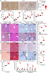
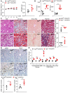
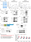
Background: Impaired mitochondrial function contributes to non-alcoholic steatohepatitis (NASH). Acylglycerol kinase (AGK) is a subunit of the translocase of the mitochondrial inner membrane 22 (TIM22) protein import complex. AGK mutation is the leading cause of Sengers syndrome, characterized by congenital cataracts, hypertrophic cardiomyopathy, skeletal myopathy, lactic acidosis, and liver dysfunction. The potential roles and mechanisms of AGK in NASH are not yet elucidated. Methods: Hepatic-specific AGK-deficient mice and AGK G126E mutation (AGK kinase activity arrest) mice were on a choline-deficient and high-fat diet (CDAHFD) and a methionine choline-deficient diet (MCD). The mitochondrial function and the molecular mechanisms underlying AGK were investigated in the pathogenesis of NASH. Results: The levels of AGK were significantly downregulated in human NASH liver samples. AGK deficiency led to severe liver damage and lipid accumulation in mice. Aged mice lacking hepatocyte AGK spontaneously developed NASH. AGK G126E mutation did not affect the structure and function of hepatocytes. AGK deficiency, but not AGK G126E mice, aggravated CDAHFD- and MCD-induced NASH symptoms. AGK deficiency-induced liver damage could be attributed to hepatic mitochondrial dysfunction. The mechanism revealed that AGK interacts with mitochondrial respiratory chain complex I subunits, NDUFS2 and NDUFA10, and regulates mitochondrial fatty acid metabolism. Moreover, the AGK DGK domain might directly interact with NDUFS2 and NDUFA10 to maintain the hepatic mitochondrial respiratory chain complex I function. Conclusions: The current study revealed the critical roles of AGK in NASH. AGK interacts with mitochondrial respiratory chain complex I to maintain mitochondrial integrity via the kinase-independent pathway.
Keywords: NDUFS2; fatty acid metabolism; mitochondrial ROS; mitochondrial respiratory chain.
© The author(s).
Conflict of interest statement
Competing Interests: The authors have declared that no competing interest exists.
Figures


Similar articles
-
Sengers Syndrome-Associated Mitochondrial Acylglycerol Kinase Is a Subunit of the Human TIM22 Protein Import Complex.Mol Cell. 2017 Aug 3;67(3):457-470.e5. doi: 10.1016/j.molcel.2017.06.014. Epub 2017 Jul 14. Mol Cell. 2017. PMID: 28712726
-
Acylglycerol Kinase Mutated in Sengers Syndrome Is a Subunit of the TIM22 Protein Translocase in Mitochondria.Mol Cell. 2017 Aug 3;67(3):471-483.e7. doi: 10.1016/j.molcel.2017.06.013. Epub 2017 Jul 14. Mol Cell. 2017. PMID: 28712724
-
Characterization of a Novel Splicing Variant in Acylglycerol Kinase (AGK) Associated with Fatal Sengers Syndrome.Int J Mol Sci. 2021 Dec 15;22(24):13484. doi: 10.3390/ijms222413484. Int J Mol Sci. 2021. PMID: 34948281 Free PMC article.
-
A Comparison of the Gene Expression Profiles of Non-Alcoholic Fatty Liver Disease between Animal Models of a High-Fat Diet and Methionine-Choline-Deficient Diet.Molecules. 2022 Jan 27;27(3):858. doi: 10.3390/molecules27030858. Molecules. 2022. PMID: 35164140 Free PMC article. Review.
-
Sengers syndrome: six novel AGK mutations in seven new families and review of the phenotypic and mutational spectrum of 29 patients.Orphanet J Rare Dis. 2014 Aug 20;9:119. doi: 10.1186/s13023-014-0119-3. Orphanet J Rare Dis. 2014. PMID: 25208612 Free PMC article. Review.
Cited by
-
ACACA reduces lipid accumulation through dual regulation of lipid metabolism and mitochondrial function via AMPK- PPARα- CPT1A axis.J Transl Med. 2024 Feb 23;22(1):196. doi: 10.1186/s12967-024-04942-0. J Transl Med. 2024. PMID: 38395901 Free PMC article.
-
Global burden of alcoholic cardiomyopathy in middle-aged men (1990-2021) and projections to 2050: a systematic analysis of the Global Burden of Disease 2021 data.Front Public Health. 2025 Jun 26;13:1608351. doi: 10.3389/fpubh.2025.1608351. eCollection 2025. Front Public Health. 2025. PMID: 40642236 Free PMC article.
-
CAV3 alleviates diabetic cardiomyopathy via inhibiting NDUFA10-mediated mitochondrial dysfunction.J Transl Med. 2024 Apr 26;22(1):390. doi: 10.1186/s12967-024-05223-6. J Transl Med. 2024. PMID: 38671439 Free PMC article.
-
ZDHHC2-Mediated AGK Palmitoylation Activates AKT-mTOR Signaling to Reduce Sunitinib Sensitivity in Renal Cell Carcinoma.Cancer Res. 2023 Jun 15;83(12):2034-2051. doi: 10.1158/0008-5472.CAN-22-3105. Cancer Res. 2023. PMID: 37078777 Free PMC article.
-
Gut dysbiosis in nonalcoholic fatty liver disease: pathogenesis, diagnosis, and therapeutic implications.Front Cell Infect Microbiol. 2022 Nov 8;12:997018. doi: 10.3389/fcimb.2022.997018. eCollection 2022. Front Cell Infect Microbiol. 2022. PMID: 36425787 Free PMC article. Review.
References
-
- Rinella M, Charlton M. The globalization of nonalcoholic fatty liver disease: Prevalence and impact on world health. Hepatology. 2016;64:19–22. - PubMed
-
- Younossi ZM, Koenig AB, Abdelatif D, Fazel Y, Henry L, Wymer M. Global epidemiology of nonalcoholic fatty liver disease-Meta-analytic assessment of prevalence, incidence, and outcomes. Hepatology. 2016;64:73–84. - PubMed
-
- Perez-Carreras M, Del Hoyo P, Martin MA, Rubio JC, Martin A, Castellano G. et al. Defective hepatic mitochondrial respiratory chain in patients with nonalcoholic steatohepatitis. Hepatology. 2003;38:999–1007. - PubMed
Publication types
MeSH terms
Substances
LinkOut - more resources
Full Text Sources
Medical

