

Human Dystrophin Dp71ab Enhances the Proliferation of Myoblasts Across Species But Not Human Nonmyoblast Cells
- PMID: 35547811
- PMCID: PMC9081641
- DOI: 10.3389/fcell.2022.877612
Human Dystrophin Dp71ab Enhances the Proliferation of Myoblasts Across Species But Not Human Nonmyoblast Cells
Abstract
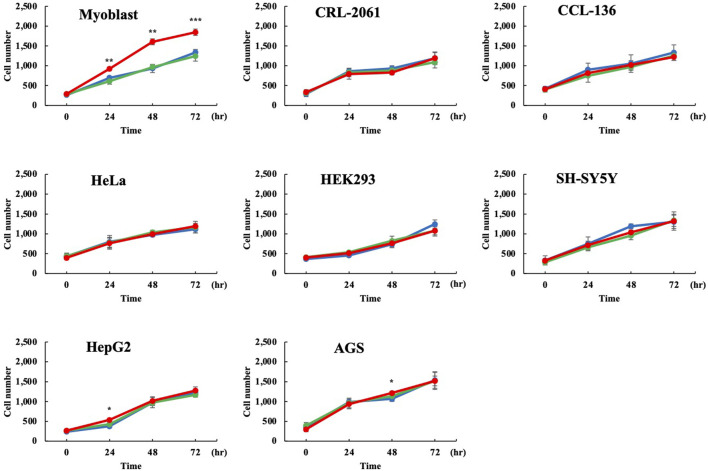
Dystrophin Dp71 is an isoform produced from the Dp71 promoter in intron 62 of the DMD gene, mutations in which cause Duchenne muscular dystrophy. Dp71 is involved in various cellular processes and comprises more than 10 isoforms produced by alternative splicing. Dp71ab, in which both exons 71 and 78 are deleted, has a hydrophobic C-terminus that is hydrophilic in Dp71. Therefore, Dp71ab is believed to have different roles from Dp71. Previously, we reported that Dp71ab enhanced the proliferation of human myoblasts. Here, we further characterized Dp71ab, focusing on the activation of cell proliferation. Dp71ab increased the proliferation of immortalized human myoblasts in a dose-dependent manner. In contrast, Dp71 suppressed proliferation in a dose-dependent manner. Consistent with these opposite effects, eGFP-tagged Dp71ab and mCherry-tagged Dp71 showed different cellular distributions, with Dp71ab mostly in the nucleus. Notably, human Dp71ab enhanced the proliferation of rat and mouse myoblasts. Despite these findings, human Dp71ab did not enhance the proliferation of human nonmyoblast cells, including rhabdomyosarcoma cells. We concluded that Dp71ab is a myoblast-specific proliferation enhancer. In further studies, Dp71ab will be employed for the expansion of myoblasts in clinical settings.
Keywords: DMD; Dp71; Dp71ab; cell proliferation; dystrophin; isoform; myoblast.
Copyright © 2022 Farea, Maeta, Nishio and Matsuo.
Conflict of interest statement
KM is employed by KNC Laboratories Co., Ltd., Kobe, Japan. MM discloses being employed by Kobe Gakuin University, which received funding from KNC Laboratories Co., Ltd., Kobe, Japan. MM further discloses being a scientific adviser for Daiichi-Sankyo Co, Tokyo, Japan and JCR Pharma Co, Ashiya, Japan. The remaining authors declare that the research was conducted in the absence of any commercial or financial relationships that could be construed as a potential conflict of interest.
Figures


Similar articles
-
Dystrophin Dp71ab is monoclonally expressed in human satellite cells and enhances proliferation of myoblast cells.Sci Rep. 2020 Oct 13;10(1):17123. doi: 10.1038/s41598-020-74157-y. Sci Rep. 2020. PMID: 33051488 Free PMC article.
-
HEK293 cells express dystrophin Dp71 with nucleus-specific localization of Dp71ab.Histochem Cell Biol. 2016 Sep;146(3):301-9. doi: 10.1007/s00418-016-1439-2. Epub 2016 Apr 25. Histochem Cell Biol. 2016. PMID: 27109495
-
Identification of the shortest splice variant of Dp71, together with five known variants, in glioblastoma cells.Biochem Biophys Res Commun. 2019 Jan 8;508(2):640-645. doi: 10.1016/j.bbrc.2018.11.168. Epub 2018 Dec 5. Biochem Biophys Res Commun. 2019. PMID: 30527806
-
Dystrophin Dp71: the smallest but multifunctional product of the Duchenne muscular dystrophy gene.Mol Neurobiol. 2012 Feb;45(1):43-60. doi: 10.1007/s12035-011-8218-9. Epub 2011 Nov 22. Mol Neurobiol. 2012. PMID: 22105192 Review.
-
Combining genetics, neuropsychology and neuroimaging to improve understanding of brain involvement in Duchenne muscular dystrophy - a narrative review.Neuromuscul Disord. 2020 Jun;30(6):437-442. doi: 10.1016/j.nmd.2020.05.001. Epub 2020 May 16. Neuromuscul Disord. 2020. PMID: 32522501 Review.
Cited by
-
CRISPR-Cas9 correction in the DMD mouse model is accompanied by upregulation of Dp71f protein.Mol Ther Methods Clin Dev. 2023 Jun 17;30:161-180. doi: 10.1016/j.omtm.2023.06.006. eCollection 2023 Sep 14. Mol Ther Methods Clin Dev. 2023. PMID: 37457303 Free PMC article.
-
Nuclear Small Dystrophin Isoforms during Muscle Differentiation.Life (Basel). 2023 Jun 11;13(6):1367. doi: 10.3390/life13061367. Life (Basel). 2023. PMID: 37374149 Free PMC article.
-
Duchenne Muscular Dystrophy from Brain to Muscle: The Role of Brain Dystrophin Isoforms in Motor Functions.J Clin Med. 2023 Aug 29;12(17):5637. doi: 10.3390/jcm12175637. J Clin Med. 2023. PMID: 37685704 Free PMC article.
References
-
- Aragon J., Martínez-Herrera A., Romo-Yáñez J., Ceja V., Azotla-Vilchis C., Siqueiros-Márquez L., et al. (2016). Identification of Dp71 Isoforms Expressed in PC12 Cells: Subcellular Localization and Colocalization with β-Dystroglycan and α1-Syntrophin. J. Mol. Neurosci. 58, 201–209. 10.1007/s12031-015-0657-8 - DOI - PubMed
-
- Aragon J., González-Reyes M., Romo-Yáñez J., Vacca O., Aguilar-González G., Rendón A., et al. (2018). Dystrophin Dp71 Isoforms Are Differentially Expressed in the Mouse Brain and Retina: Report of New Alternative Splicing and a Novel Nomenclature for Dp71 Isoforms. Mol. Neurobiol. 55, 1376–1386. 10.1007/s12035-017-0405-x - DOI - PubMed
LinkOut - more resources
Full Text Sources