[Preprint]. 2023 Jan 19:2021.09.15.460543.
doi: 10.1101/2021.09.15.460543.

Dual domain recognition determines SARS-CoV-2 PLpro selectivity for human ISG15 and K48-linked di-ubiquitin

Affiliations

Dual domain recognition determines SARS-CoV-2 PLpro selectivity for human ISG15 and K48-linked di-ubiquitin

Pawel M Wydorski et al. bioRxiv. .

Update in

Abstract

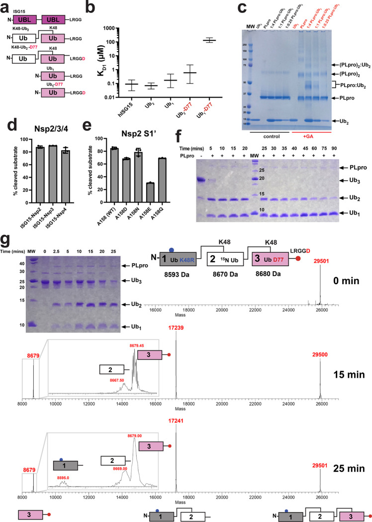
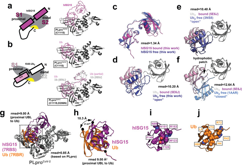
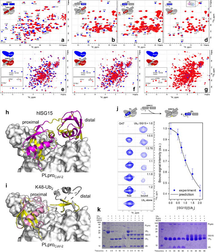
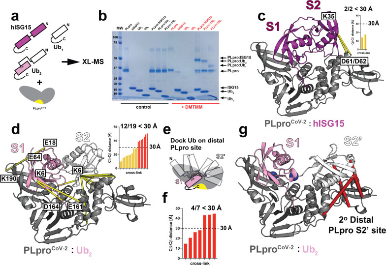
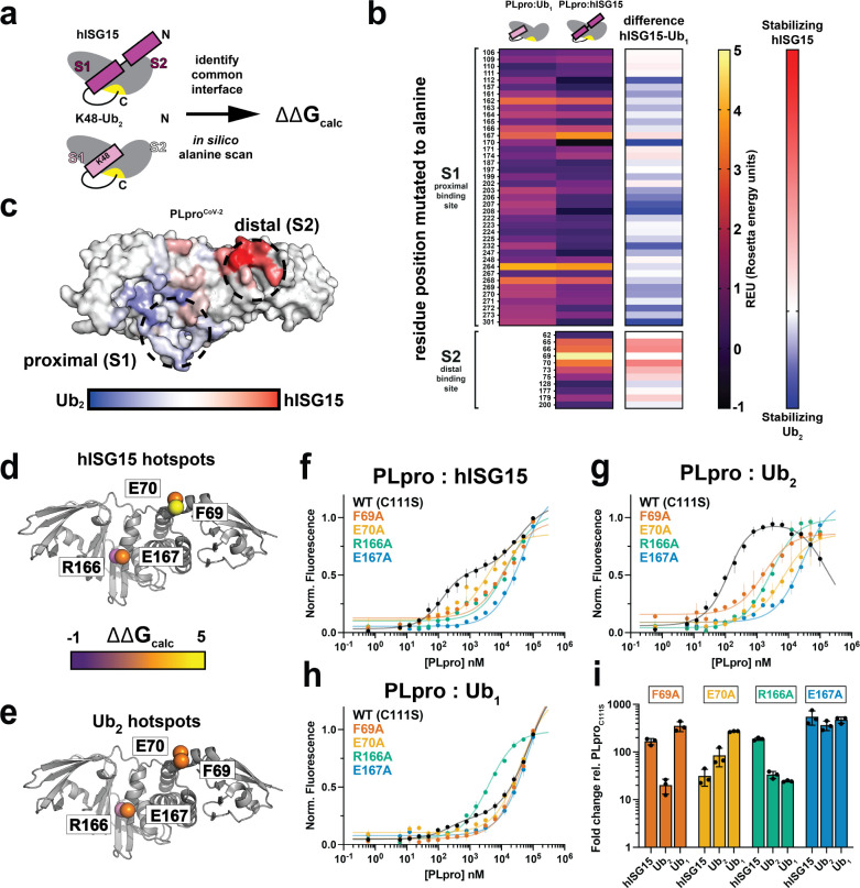
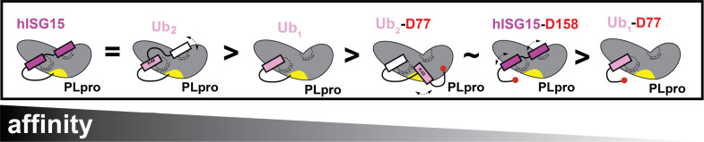
The Papain-like protease (PLpro) is a domain of a multi-functional, non-structural protein 3 of coronaviruses. PLpro cleaves viral polyproteins and posttranslational conjugates with poly-ubiquitin and protective ISG15, composed of two ubiquitin-like (UBL) domains. Across coronaviruses, PLpro showed divergent selectivity for recognition and cleavage of posttranslational conjugates despite sequence conservation. We show that SARS-CoV-2 PLpro binds human ISG15 and K48-linked di-ubiquitin (K48-Ub 2 ) with nanomolar affinity and detect alternate weaker-binding modes. Crystal structures of untethered PLpro complexes with ISG15 and K48-Ub 2 combined with solution NMR and cross-linking mass spectrometry revealed how the two domains of ISG15 or K48-Ub 2 are differently utilized in interactions with PLpro. Analysis of protein interface energetics predicted differential binding stabilities of the two UBL/Ub domains that were validated experimentally. We emphasize how substrate recognition can be tuned to cleave specifically ISG15 or K48-Ub 2 modifications while retaining capacity to cleave mono-Ub conjugates. These results highlight alternative druggable surfaces that would inhibit PLpro function.


Conflict of interest statement

COMPETING INTEREST

The authors declare that they have no competing interests.

Figures

Figure 1.
Figure 1.. PLproCoV-2 substrate binding and recognition.
(a) Topology of utilized substrates: ISG15, K48-linked Ub2 ending with G76 on the C-terminus (K48-Ub2), K48-linked Ub2 ending with D77 on the C-terminus (K48-Ub2-D77), and corresponding monomeric ubiquitins. (b) MST binding analysis of substrates to PLproCoV-2. Comparison of dissociation constants for the primary binding event (Kd1). The Kd1 is derived from a fit to three independent experiments with error bars corresponding to 68.3% confidence interval derived from error-surface projections. (c) Inter- and intramolecular non-specific cross-linking of PLproCoV-2 complexes. Cross-linked samples with different molar ratios reveal formation of heterogeneous covalent PLproCoV-2:Ub2 heterodimer complex bands (red, for glutaraldehyde cross-linker) compared to untreated reactions (black, control) by SDS-PAGE. (d) Quantification of the cleavage efficiency of hISG15 C-terminal fusions with peptides from Nsp2 (AYTRYVDNNF), Nsp3 (APTKVTFGDD), and Nsp4 (KIVNNWLKQL) from SARS-CoV-2 that mimic natural substrates of PLproCoV-2, as revealed by SDS-PAGE gel. Shown is the percentage of the input population that has been cut by PLproCoV-2. Experiment was performed in triplicate and is reported as an average with standard deviation. (e) Quantification of the cleavage efficiency of Nsp2 peptides as dependent on amino acid located at the C-terminus of “LRGG(X)” motif. Shown is the percentage of the input population that has been cut by PLproCoV-2. Experiment was performed in triplicate and is reported as an average with standard deviation. (f) PLproCoV-2 cleavage of K48-Ub3. SDS-PAGE gel reveals that PLpro cleaves Ub3 into Ub2 and Ub1 efficiently with various rates. (g) Mass spectrometry detection of cleavage patterns for PLproCoV-2 hydrolyzing Ub3, in which the distal Ub (1) carries K48R mutation, the endo (2) Ub is 15N-labeled, and the proximal (3) Ub contains C-terminal D77 extension. Analysis of the time course reveals that this Ub3 is primarily hydrolyzed between Ubs 2 and 3. Masses of individual Ub units are shown on the top, and the identified products are shown at the bottom.
Figure 2.
Figure 2.. MX structure of PLproCoV-2 bound to human ISG15 and K48-Ub2 reveals differential usage of distal domains.
Schematic of PLpro bound to (a, left) hISG15 and (b, left) K48-linked Ub2. PLpro is shown in gray with an active site indicated in yellow. hISG15 is shown in magenta. Proximal and distal Ubs are pink and white, respectively. Crystal structures of PLproCoV-2 bound to (a, right) hISG15 and (b, right) K48-Ub2. PLpro (C111S or C111S,D286N) is shown in cartoon representation, hISG15 and K48-Ub2 are shown as a backbone trace. PLpro, hISG15 and K48-Ub2 are colored as in (a, left) and (b, left). (c) Overlay of crystal structures of bound hISG15 (magenta, PDB id: 7RBS, this study) and unbound hISG15 (blue, PDB id: 7S6P, this study). (d) Overlay of bound conformation of K48-linked Ub2 observed in complex with PLproCoV-1 (PDB id: 5E6J) with unbound conformation of K48-linked Ub2 (PDB id: 7S6O, this study). Proximal Ub of both bound and unbound conformations is shown in pink, distal Ub of bound conformation is shown in white, and distal Ub of unbound conformation is shown in blue. (e) Overlay of bound conformation of K48-linked Ub2 observed in complex with PLproCoV-1 (PDB id: 5E6J) with unbound “open” conformation of Ub2 (PDB id: 3NS8). Ub units are shown as in (d). (f) Overlay of bound conformation of K48-linked Ub2 observed in complex with PLproCoV-1 (PDB id: 5E6J) with unbound “closed” conformation of Ub2 (PDB id: 1AAR). Ub units are shown as in (d). Intramolecular Ub-Ub interface is indicated with an arrow. (g) Structural overlay of PLproCoV-2:hISG15 and PLproCoV-2:Ub2 (PDB id: 7RBR, this study). Proteins are shown in cartoon and colored gray (PLpro), magenta (hISG15) and orange (Ub2). (h) Zoom in of the boxed area in (g) representing overlay of the proximal domain of hISG15 and Ub. Proximal domain of hISG15 and Ub are represented as in (g). Rotation of the binding surface indicated with arrows. (i,j) Comparison of the binding surfaces of hISG15 (i) and Ub1 (j). Proximal domain of hISG15 and Ub are represented as in (g). Key residues are shown in space fill representation.
Figure 3.
Figure 3.. NMR data showing PLproCoV-2 interactions with hISG15 and K48-Ub2.
(a-d) Overlay of 1H-15N SOFAST-HMQC spectra of 15N-labeled (a) hISG15, (b) distal Ub in Ub2, (c) proximal Ub in Ub2, and (d) monomeric Ub, alone (blue) and with 1.2 molar equivalents of unlabeled PLproCoV-2 (C111S) (red). Signals of select residues are indicated. The inset in d illustrates gradual shift of G47 signal during titration. (e-g) Overlay of 1H-15N TROSY spectra of 15N-PLproCoV-2 (C111S,Y171H) alone (blue) and (in red) with 1.5 molar equivalents of unlabeled (e) hISG15, (f) Ub2, and (g) monomeric Ub (red). Insets zoom on the region containing indole HN signals of tryptophans (W93 and W106) and HN of imidazole ring of histidine H272. (i, top panel) hISG15 residues with strong signal perturbations mapped (yellow) on our structure of PLproCoV-2:hISG15 complex. (i, bottom panel) Residues in the proximal and distal Ubs with strong signal perturbations mapped (yellow) on our structure of PLproCoV-2:K48-Ub2 complex. (j) Competition for PLproCoV-2 binding between Ub2 and ISG15. Shown on the left are representative 1H-15N NMR signals of G47 in 15N-labeled proximal Ub of Ub2 mixed with unlabeled PLproCoV-2 (C111S,Y171H) (in 1:1.5 molar ratio) upon addition of unlabeled hISG15, for indicated values of hISG15:Ub2 molar ratio. Shown on the right is the intensity of the PLpro-bound signal of G47 as a function of [hISG15]:[Ub2] (dots) and the predicted molar fraction of bound Ub2 (line) using the Kd1 values obtained in this work (Supplementary Table 1). (k) SDS-PAGE gels showing the inhibitory effect of hISG15 on disassembly of K48-linked Ub2 by PLproCoV-2 (right gel) and minimal effect (if any) of hISG15 on disassembly of K48-linked Ub3 (left gel). The hISG15 and Ub2 constructs used here all had G (G157 or G76) as the C-terminal residue.
Figure 4.
Figure 4.. Cross-linking mass spectrometry (XL-MS) analysis of PLpro in complex with hISG15 and K48-Ub2.
(a) Schematic illustration of cross-linking mass spectrometry experiments for heterodimer complexes of PLproCoV-2 with hISG15, K48-Ub2, and Ub1. Active site of PLproCoV-2 is indicated in yellow. (b) Cross-linked samples reveal formation of covalent heterodimer complex bands (red, DMTMM cross-linker) compared to untreated reactions (black, control) by SDS-PAGE. (c) Interactions between PLproCoV-2 (D61/D62) and the distal hISG15 domain (K35) identified by XL-MS mapped on the structure of heterocomplex. Both identified contacts are shorter than 30 Å. PLpro (gray) and hISG15 (magenta) are shown in cartoon representation. Cross-links are colored yellow. (d) Interactions between PLproCoV-2 and K48-Ub2 identified by XL-MS mapped on the structure of the heterocomplex. Twelve contacts found to be shorter than 30 Å show interaction of K48-Ub2 with fingers, palm and thumb domains of PLproCoV-2. PLproCoV-2 is shown in cartoon representation and colored gray. K48-Ub2 is shown in cartoon representation and colored white and pink for the proximal and distal Ubs, respectively. Cross-links are colored yellow. (e) Modeling strategy showing docked Ub monomer to PLproCoV-2:Ub complex with Ub placed into the S1 binding site. Utilized constraint maintaining proximity between K48 of the proximal Ub and C-terminus of the docked Ub is shown in blue spheres. (f) A low energy model generated with strategy presented in (e) explains 4 of the remaining 7 contacts from the XL-MS in (d). (g) Contacts shorter than 30 Å between low energy model of PLproCoV-2 bound to Ub2 in the S1 and S2’ sites as shown in (e, f). Cross-links between distal Ub and thumb and UBL domains of PLproCoV-2 are colored red. Constraint used in docking (e) is indicated as blue spheres. PLproCoV-2 and K48-Ub2 are shown as in (d).
Figure 5.
Figure 5.. Prediction and validation of specificity determining surfaces on PLproCoV-2.
(a) Schematic illustration of identification of substrate binding surfaces on PLproCoV-2 and in silico mutagenesis for heterodimer complexes with hISG15 and K48-Ub2. (b) Heat-map results of ΔΔGbinding calculations of in silico alanine scan for PLproCoV-2 in complex with hISG15 or Ub1. Interface residue positions in the S1 (proximal) and S2 (distal) binding sites are labeled. The heat-map is colored from black to yellow. The last column represents results of calculations of difference between REU (Rosetta energy units) for PLproCoV-2:hISG15 and PLproCoV-2:K48-Ub2 and is colored from blue to red. (c) Results of in silico mutagenesis for complexes of PLproCoV-2 with hISG15 and K48-Ub2 mapped on surface representation of PLproCoV-2. Hotspot sites identified as those driving stability towards hISG15 are colored red, and those driving stability towards K48-Ub2 are colored blue. Summary of PLproCoV-2 alanine mutants (F69A, E70A. R166A and E167A) tested for binding to hISG15 (d), Ub2 (e) and Ub1. Mutants are shown as Ca spheres and are colored according to ΔΔGcalc from black to yellow. (f-h) PLproCoV-2 WT (C111S) (black), PLproCoV-2 F69A (orange), PLproCoV-2 E70A (yellow), PLproCoV-2 R166A (green) and PLproCoV-2 E167A (blue) titrations with hISG15 (f), Ub2 (g) and Ub1 (h). Data is shown as triplicates and is plotted as the average with the range of individual replicates. Data is fitted to the preferred 1:2 binding model for WT (C111S) and to 1:1 binding model for mutants using PALMIST. (i) Summary of fold change in Kds calculated as a ratio between PLproCoV-2 WT (C111S) and PLproCoV-2 F69A, PLproCoV-2 E70A, PLproCoV-2 R166A, PLproCoV-2 E167A to hISG15, Ub2 and Ub1. Data is shown as triplicates and is plotted as averages with standard deviation.
Figure 6.
Figure 6.. Dual domain-based model for PLpro recognition of K48-linked Ub2 and ISG15.
Schematic representation of differences in binding of PLproCoV-2 with hISG15 and ubiquitin species.

Similar articles

References

    1. Kim D. et al. A high-resolution temporal atlas of the SARS-CoV-2 translatome and transcriptome. Nat Commun 12, 5120 (2021). - PMC - PubMed
    1. Kim D. et al. The Architecture of SARS-CoV-2 Transcriptome. Cell 181, 914–921.e10 (2020). - PMC - PubMed
    1. Mahmoudvand S. & Shokri S. Interactions between SARS coronavirus 2 papain-like protease and immune system: A potential drug target for the treatment of COVID-19. Scand J Immunol 94, (2021). - PMC - PubMed
    1. Ziebuhr J., Thiel V. & Gorbalenya A. E. The Autocatalytic Release of a Putative RNA Virus Transcription Factor from Its Polyprotein Precursor Involves Two Paralogous Papain-like Proteases That Cleave the Same Peptide Bond. Journal of Biological Chemistry 276, 33220–33232 (2001). - PMC - PubMed
    1. Mielech A. M., Chen Y., Mesecar A. D. & Baker S. C. Nidovirus papain-like proteases: Multifunctional enzymes with protease, deubiquitinating and deISGylating activities. Virus Research 194, 184–190 (2014). - PMC - PubMed

Publication types

LinkOut - more resources