. 2018 Oct 15;8(62):35386-35394.
doi: 10.1039/c8ra06274a.

Antibacterial properties of electrospun Ti3C2T z (MXene)/chitosan nanofibers

Affiliations

Antibacterial properties of electrospun Ti3C2T z (MXene)/chitosan nanofibers

Elisa A Mayerberger et al. RSC Adv. .

Abstract

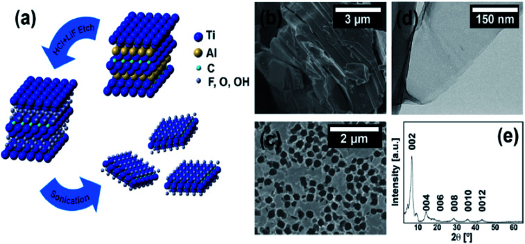
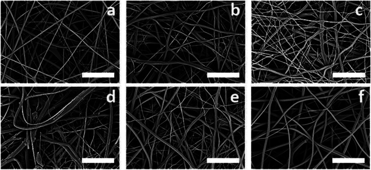
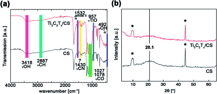
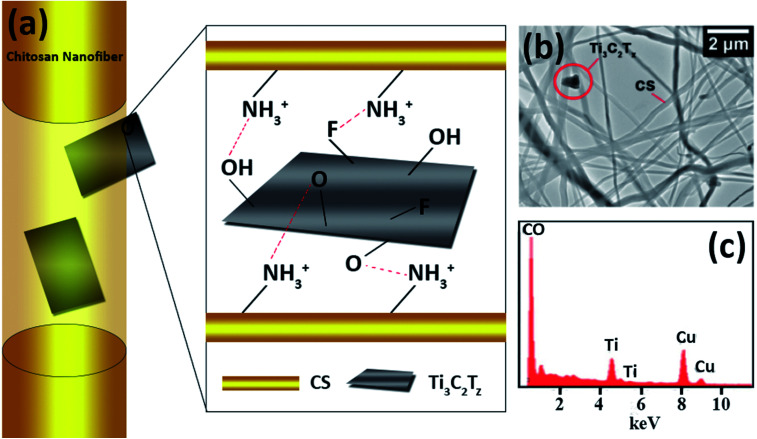
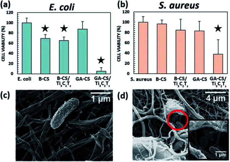
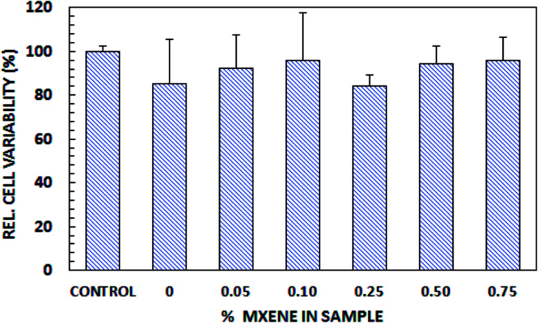
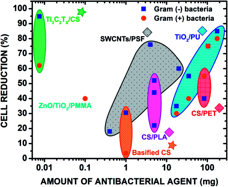
Electrospun natural polymeric bandages are highly desirable due to their low-cost, biodegradability, non-toxicity and antimicrobial properties. Functionalization of these nanofibrous mats with two-dimensional nanomaterials is an attractive strategy to enhance the antibacterial effects. Herein, we demonstrate an electrospinning process to produce encapsulated delaminated Ti3C2T z (MXene) flakes within chitosan nanofibers for passive antibacterial wound dressing applications. In vitro antibacterial studies were performed on crosslinked Ti3C2T z /chitosan composite fibers against Gram-negative Escherichia coli (E. coli) and Gram-positive Staphylococcus aureus (S. aureus) - demonstrating a 95% and 62% reduction in colony forming units, respectively, following 4 h of treatment with the 0.75 wt% Ti3C2T z - loaded nanofibers. Cytotoxicity studies to determine biocompatibility of the nanofibers indicated the antibacterial MXene/chitosan nanofibers are non-toxic. The incorporation of Ti3C2T z single flakes on fiber morphology was analyzed by scanning electron microscopy (SEM) and transmission electron microscopy equipped with an energy-dispersive detector (TEM-EDS). Our results suggest that the electrospun Ti3C2T z /chitosan nanofibers are a promising candidate material in wound healing applications.


Conflict of interest statement

There are no conflicts to declare.

Figures

Fig. 1
Fig. 1. (a) Schematic detailing the removal of the Al layers from the Ti3AlC2 to yield ML-Ti3C2Tz that washing, and sonication then separate into individual Ti3C2Tz flakes. (b) SEM micrographs of ML-Ti3C2Tz powder and, (c) Ti3C2Tz single and few-layer thick flakes on an anodized alumina membrane. (d) TEM micrograph of a Ti3C2Tz flake. (e) XRD diffractogram of the vacuum assisted filtered Ti3C2Tz film.
Fig. 2
Fig. 2. SEM micrographs displaying the surface of the as-spun electrospun crosslinked Ti3C2Tz/CS mats for the five different MXene loadings: (a) 0, (b) 0.05, (c) 0.10, (d) 0.25, (e) 0.50, and (f) 0.75 wt%. Scale bars represent 5 μm.
Fig. 3
Fig. 3. (a) FTIR spectra and (b) XRD patterns of the neat chitosan nanofibers and the 0.75 wt% Ti3C2Tz/CS nanofibers. Asterisks denote signal by the stage.
Fig. 4
Fig. 4. (a) Schematic diagram illustrating the observed orientations of Ti3C2Tz flakes within the chitosan nanofibers. (b) TEM micrograph of the Ti3C2Tz/CS nanofibers and (c) elemental analysis (the presence of copper arises from the TEM grid).
Fig. 5
Fig. 5. Antibacterial activity of, (a) E. coli and (b) S. aureus. B-X and GA-X indicate mats treated with NaOH and glutaraldehyde, respectively. SEM micrographs shows an, (c) intact and, (d) destroyed E. coli bacteria on the 0.75 wt% Ti3C2Tz/CS nanofiber mat. The star icon designates samples that are significantly different from the control, p ≤ 0.05.
Fig. 6
Fig. 6. Biocompatibility of Ti3C2Tz/CS nanofibers.
Fig. 7
Fig. 7. Comparison of the antibacterial properties with the previous literature of select polymeric nanofibers with incorporated metal oxides and 2D materials. The antibacterial effects of electrospun polymer blends were also compared. The stars denote the materials from this study. Copyright WILEY-VCH Verlag GmbH & Co. KGaA, 69469 Weinheim, Germany, 2016.

Similar articles

Cited by

References

    1. Bitounis D. Ali-Boucetta H. Hong B. H. Min D.-H. Kostarelos K. Adv. Mater. 2013;25:2258–2268. doi: 10.1002/adma.201203700. - DOI - PubMed
    1. Chung C. Kim Y.-K. Shin D. Ryoo S.-R. Hong B. H. Min D.-H. Acc. Chem. Res. 2013;46:2211–2224. doi: 10.1021/ar300159f. - DOI - PubMed
    1. Gurunathan S. Han J. W. Dayem A. A. Eppakayala V. Kim J.-H. Int. J. Nanomed. 2012;7:5901. doi: 10.2147/IJN.S37397. - DOI - PMC - PubMed
    1. Zhang Y. Nayak T. R. Hong H. Cai W. Nanoscale. 2012;4:3833–3842. doi: 10.1039/C2NR31040F. - DOI - PMC - PubMed
    1. Naguib M. Mashtalir O. Carle J. Presser V. Lu J. Hultman L. Gogotsi Y. Barsoum M. W. ACS Nano. 2012;6:1322–1331. doi: 10.1021/nn204153h. - DOI - PubMed